

Safety assurance in obstetrical ultrasound
- PMID: 18450141
- PMCID: PMC2390856
- DOI: 10.1053/j.sult.2007.12.003
Safety assurance in obstetrical ultrasound
Abstract
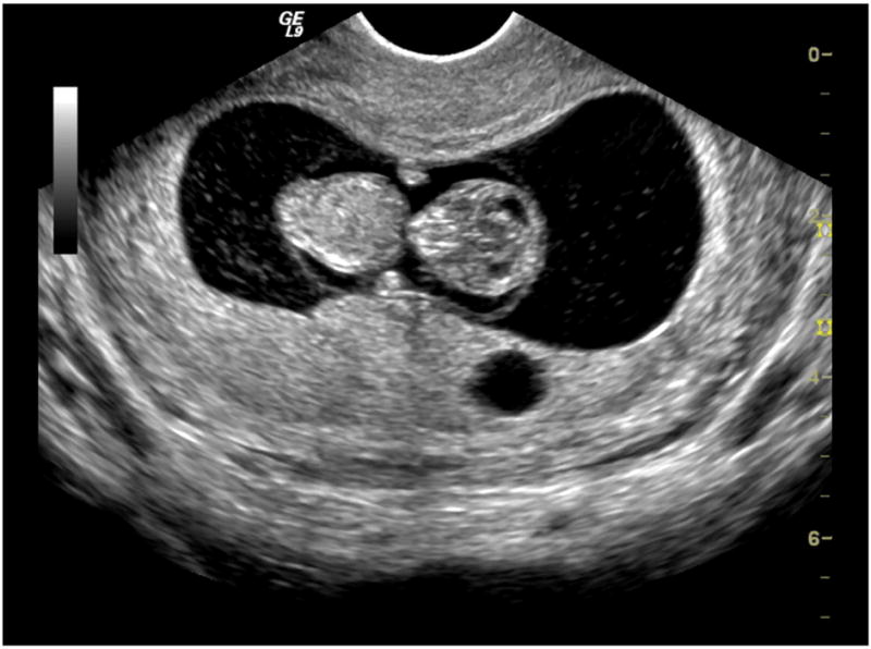
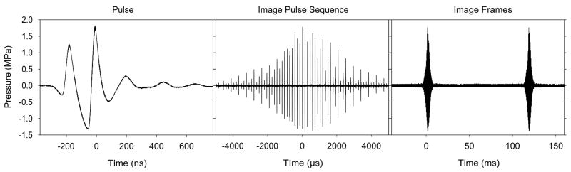
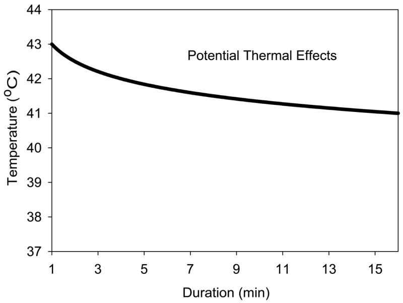
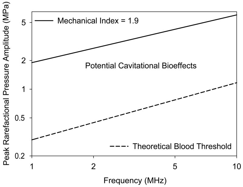
Safety assurance for diagnostic ultrasound in obstetrics began with a tacit assumption of safety allowed by a federal law enacted in 1976 for then-existing medical ultrasound equipment. The implementation of the 510(k) pre-market-approval process for diagnostic ultrasound resulted in the establishment of guideline upper limits for several examination categories in 1985. The obstetrical category has undergone substantial evolution from initial limits (ie, 46 mW/cm2 spatial peak temporal average [SPTA] intensity) set in 1985. Thermal and mechanical exposure indices, which are displayed onscreen according to an Output Display Standard, were developed for safety assurance with relaxed upper limits. In 1992, with the adoption of the Output Display Standard, the allowable output for obstetrical ultrasound was increased in terms of both the average exposure (eg, to a possible 720 mW/cm2 SPTA intensity) and the peak exposure (via the Mechanical Index). There has been little or no subsequent research with the modern obstetrical ultrasound machines to systematically assess potential risks to the fetus using either relevant animal models of obstetrical exposure or human epidemiology studies. The assurance of safety for obstetrical ultrasound therefore is supported by three ongoing means: (1) review of a substantial but uncoordinated bioeffect research literature; (2) the theoretical evaluation of diagnostic ultrasound exposure in terms of thermal and nonthermal mechanisms for bioeffects; and (3) the skill and knowledge of professional sonographers. At this time, there is no specific reason to suspect that there is any significant health risk to the fetus or mother from exposure to diagnostic ultrasound in obstetrics. This assurance of safety supports the prudent use of diagnostic ultrasound in obstetrics by trained professionals for any medically indicated examination.
Figures
References
-
- Fry WJ. Recent developments in ultrasound at the biophysical research laboratory and their application to basic problems in biology and medicine. In: Kelly E, editor. Ultrasonic Energy. Urbana, Il: University of Illinois Press; 1965. pp. 200–228.
-
- Reid JM, Joyner CR. The use of ultrasound to record the motion of heart structure. In: Kelly E, editor. Ultrasonic Energy. Urbana, Il: University of Illinois Press; 1965. pp. 200–228.
-
- Kossoff G, Garrett WJ, Robinson DE. An ultrasonic echoscope for visualizing the pregnant uterus. In: Kelly E, editor. Ultrasonic Energy. Urbana, Il: University of Illinois Press; 1965. pp. 200–228.
-
- Reid JM, Sikov MR. Interaction of Ultrasound and BIlogical Tissues. Rockville, MD: USDHEW; 1972.
-
- NIH. Diagnostic Ultrasound Imaging in Pregnancy, NIH Publication No. 84–667. Bethesda, MD: National Institutes of Health; 1984.
Publication types
MeSH terms
Grants and funding
LinkOut - more resources
Full Text Sources
