. 2008 May;28(5):621-9.
doi: 10.1016/j.immuni.2008.03.015. Epub 2008 May 1.

MicroRNA-155 is a negative regulator of activation-induced cytidine deaminase

Affiliations

MicroRNA-155 is a negative regulator of activation-induced cytidine deaminase

Grace Teng et al. Immunity. 2008 May.

Abstract

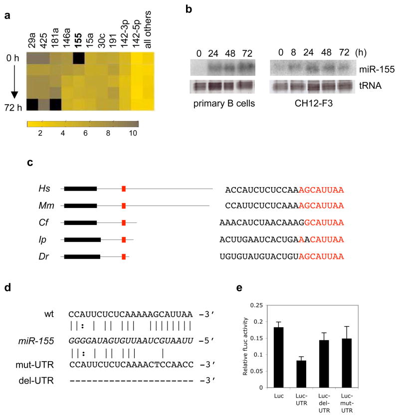
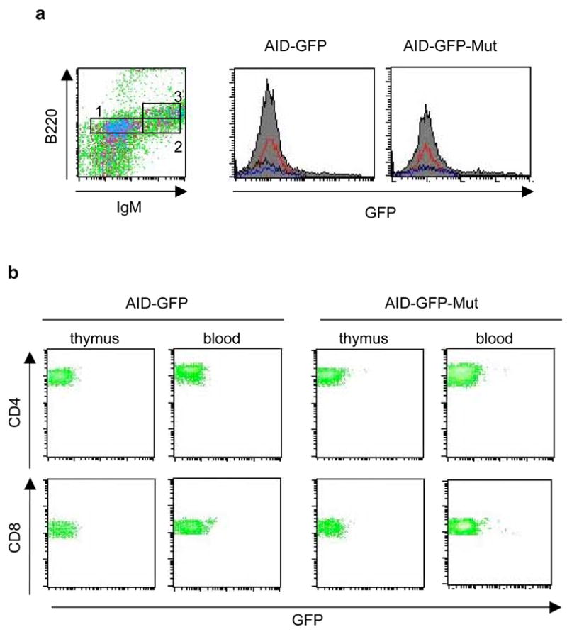
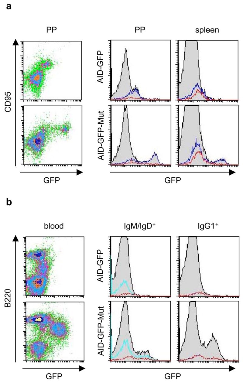
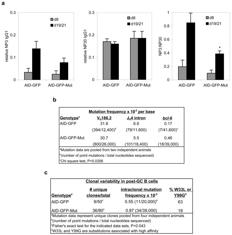
B lymphocytes perform somatic hypermutation and class-switch recombination (CSR) of the immunoglobulin locus to generate an antibody repertoire diverse in both affinity and function. These somatic diversification processes are catalyzed by activation-induced cytidine deaminase (AID), a potent DNA mutator whose expression and function are highly regulated. Here we show that AID was regulated posttranscriptionally by a lymphocyte-specific microRNA, miR-155. We found that miR-155 was upregulated in murine B lymphocytes undergoing CSR and that it targeted a conserved site in the 3'-untranslated region of the mRNA encoding AID. Disruption of this target site in vivo resulted in quantitative and temporal deregulation of AID expression, along with functional consequences for CSR and affinity maturation. Thus, miR-155, which has recently been shown to play important roles in regulating the germinal-center reaction, does so in part by directly downmodulating AID expression.


Figures

Figure 1
Figure 1
miR-155 is upregulated in B cells undergoing CSR, and targets the 3′UTR of AID. a, Relative cloning frequencies for 10 of the most abundant miRNAs in murine splenic B lymphocytes stimulated with IL-4 and LPS (0, 8, 24, 48, and 72 hours). Color coding scale is shown at bottom, and undetected miRNAs are indicated by black. b, Northern blots for miR-155 from total RNA isolated from murine splenic B cells (left) and the murine B cell line CH12-F3 (right), stimulated in vitro (0–72 hours) to undergo CSR. Ethidium-bromide staining of tRNA bands is shown as a loading control. c, Schematic of AID mRNA from human (Hs), mouse (Mm), dog (Cf), catfish (Ip), and zebrafish (Dr); showing the AID ORF (black boxes), UTRs (black lines), and the predicted miR-155 target site (red). Also shown are sequences of the corresponding miR-155 target sites, with the conserved seed region highlighted in red. d, Base pairing between murine miR-155 (italicized) and its target site in the wt AID 3′UTR. Sequences of the mutated (mut-UTR) and deleted (del-UTR) target site variants are shown underneath. e, CH12-F3 cells stimulated to undergo CSR were transfected with reporter constructs containing firefly luciferase alone (Luc), or fused to the wild-type AID 3′UTR (UTR), del-UTR, or mut-UTR. Data represent mean values of eight independent experiments ± s.e.m. One-way ANOVA F-statistic = 2.95 (P = 0.05).
Figure 2
Figure 2
Mutation of the AID miR-155 target site results in deregulated AID-GFP expression and increased CSR efficiency in vitro. a, The transgenic AID-GFP locus (which is located in the approximate center of a ~75 kbp transgene) is diagrammed, with the five coding exons of AID denoted by black boxes. The hybrid exon 5 includes the GFP gene (hatched box) inserted between the final coding portion of AID and the 3′UTR (white box). The mutated miR-155 target site is marked by an asterisk. Base pairing between miR-155 (italicized) and the wt and mutated UTRs are shown below. b, AID-GFP mRNA expression was monitored by quantitative PCR in representative AID-GFP control and AID-GFP-Mut mice. Data are normalized to Ku70 mRNA expression, and the scale is set to 1 for d0. c, AID-GFP protein expression was monitored by FACS up to four days after in vitro stimulation of splenic B lymphocytes with IL-4 and LPS. Percentages of GFP+ B220+ cells are indicated in the upper right quadrants. Cells from representative AID-GFP control (Tg copy number ~4–5) and AID-GFP-Mut (Tg copy number ~4–5) are shown. d, Median GFP fluorescence intensities, expressed in logarithmic units, are shown for the two samples in (b). e, Splenic B lymphocytes from wt (n=4); AID+/−, AID-GFP control (n=5); AID−/−, AID-GFP control (n=2); AID+/−, AID-GFP-Mut (n=6), or AID−/−, AID-GFP-Mut (n=3) mice were stimulated in vitro for three days with LPS (to induce CSR to IgG3), LPS and IL-4 (to induce CSR to IgG1), or LPS and IFNγ (to induce CSR to IgG2a). Note – AID−/−, AID-GFP control samples did not switch robustly to IgG1 or IgG3, though IgG1+ and IgG3+ populations were detected by FACS analysis(data not shown) – but they did show quantifiable levels of CSR to IgG2a. t-tests, *P <0.05, **P <0.01.
Figure 3
Figure 3
NP-immunized AID-GFP-Mut mice do not express AID-GFP in developing B lymphocytes or in T lymphocytes. FACS analyses are shown for representative AID-GFP and AID-GFP-Mut mice. a, Developing B cells (CD93+) from the bone marrow are subdivided into pro/pre-B cells (box 1 – IgM-, B220low), transitional B cells (box 2 – IgM+, B220low), and recirculating B cells (box 3 – IgM+, B220high). Histograms for GFP fluorescence show pre-pro B cells (red), transitional B cells (blue), and recirculating B cells (black). b, CD8+ and CD4+ T lymphocytes from thymus and peripheral blood are shown.
Figure 4
Figure 4
Mutation of the AID miR-155 target site results in deregulated in vivo expression of AID-GFP in NP-CGG immunized mice. a, FACS for AID-GFP expression in GC’s from representative AID-GFP and AID-GFP-Mut mice. Left, GC B cells from Peyer’s Patches (gated on B220+ B cells). Right, histograms show AID-GFP expression in B cell subsets from GC’s of Peyer’s patches (PP) and spleen. Shown in overlay are CD95+ B220+ GC B cells (blue), and the subset of GC B cells that recognize NP (red). b, FACS for AID-GFP expression in peripheral blood from representative AID-GFP and AID-GFP-Mut mice. Left, Total blood lymphocytes are shown. Right, Histograms show AID-GFP expression in IgM/IgD+, B220+ (cyan) and IgG1+, B220+ B cells (blue) subsets. Red overlays indicate the respective subsets of these cells that recognize NP (note – the blue and red overlays in the IgG1+ histogram overlap completely).
Figure 5
Figure 5
AID-GFP-Mut mice show loss of affinity maturation and increased clonal heterogeneity in post-GC B cells. a, Measurement of affinity maturation of NP-specific IgG1 by ELISA. Shown from left to right are average titers of NP3-binding IgG1, NP30-binding IgG1, and NP3:NP30-binding ratios as measured by ELISA in AID-GFP (n=3) and AID-GFP-Mut (n=7) mice ± s.e.m. t-test *P=0.0016. b, Mutation frequencies in VH186.2, JH4 intron, and bcl-6 from splenic GC B cells in NP-immunized AID-GFP and AID-GFP-Mut mice. Mutation frequencies were calculated by dividing the accumulated number of mutations in a given region by the total number of nucleotides sequenced from that region. c, Clonal heterogeneity in the JH4 intron of lymphocytes from peripheral blood of AID-GFP and AID-GFP-Mut mice. We thank Sebastian Fugmann, David Schatz, and Svend Petersen-Mahrt for comments on the manuscript. This work was supported by grants from the Keck foundation, NIH grant CA098495 (FNP), NIH NRSA training grant GM066699 (GT), and the Intramural Research Program of the National Institute of Arthritis and Musculoskeletal and Skin Diseases of the National Institutes of Health (RC).

Comment in

References

    1. Bartel DP. MicroRNAs: genomics, biogenesis, mechanism, and function. Cell. 2004;116:281–297. - PubMed
    1. Bartel DP, Chen CZ. Micromanagers of gene expression: the potentially widespread influence of metazoan microRNAs. Nat Rev Genet. 2004;5:396–400. - PubMed
    1. Basu U, Chaudhuri J, Alpert C, Dutt S, Ranganath S, Li G, Schrum JP, Manis JP, Alt FW. The AID antibody diversification enzyme is regulated by protein kinase A phosphorylation. Nature. 2005;438:508–511. - PubMed
    1. Basu U, Chaudhuri J, Phan RT, Datta A, Alt FW. Regulation of activation induced deaminase via phosphorylation. Adv Exp Med Biol. 2007;596:129–137. - PubMed
    1. Chaudhuri J, Khuong C, Alt FW. Replication protein A interacts with AID to promote deamination of somatic hypermutation targets. Nature. 2004;430:992–998. - PubMed

Publication types

MeSH terms