. 2008 Jun 27;283(26):18167-76.
doi: 10.1074/jbc.M801327200. Epub 2008 Apr 30.

Cathepsin L is responsible for processing and activation of proheparanase through multiple cleavages of a linker segment

Affiliations

Cathepsin L is responsible for processing and activation of proheparanase through multiple cleavages of a linker segment

Ghada Abboud-Jarrous et al. J Biol Chem. .

Abstract

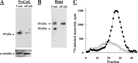
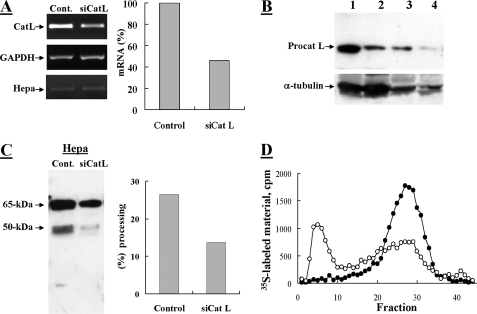
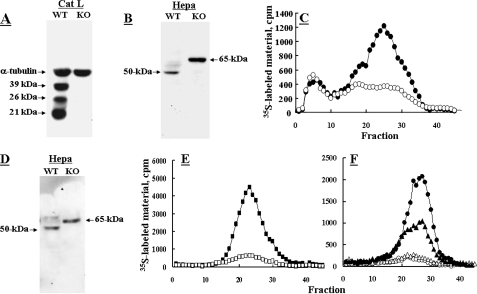
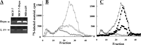
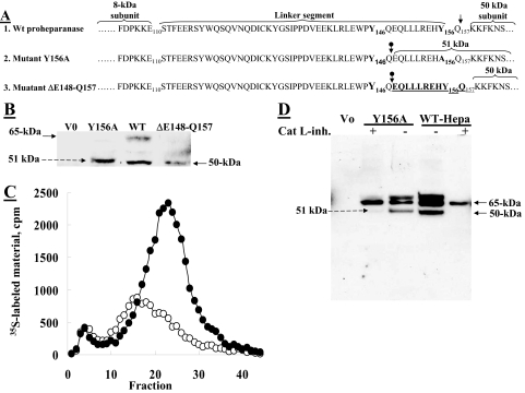
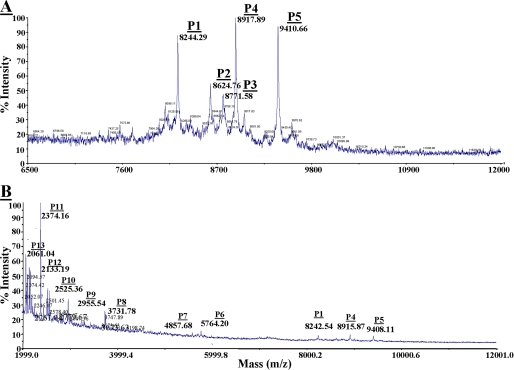
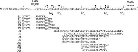
Heparanase is an endo-beta-d-glucuronidase that degrades heparan sulfate in the extracellular matrix and on the cell surface. Human proheparanase is produced as a latent protein of 543 amino acids whose activation involves excision of an internal linker segment (Ser(110)-Gln(157)), yielding the active heterodimer composed of 8- and 50-kDa subunits. Applying cathepsin L knock-out tissues and cultured fibroblasts, as well as cathepsin L gene silencing and overexpression strategies, we demonstrate, for the first time, that removal of the linker peptide and conversion of proheparanase into its active 8 + 50-kDa form is brought about predominantly by cathepsin L. Excision of a 10-amino acid peptide located at the C terminus of the linker segment between two functional cathepsin L cleavage sites (Y156Q and Y146Q) was critical for activation of proheparanase. Matrix-assisted laser desorption ionization time-of-flight mass spectrometry demonstrates that the entire linker segment is susceptible to multiple endocleavages by cathepsin L, generating small peptides. Mass spectrometry demonstrated further that an active 8-kDa subunit can be generated by several alternative adjacent endocleavages, yielding the precise 8-kDa subunit and/or slightly elongated forms. Altogether, the mode of action presented here demonstrates that processing and activation of proheparanase can be brought about solely by cathepsin L. The critical involvement of cathepsin L in proheparanase processing and activation offers new strategies for inhibiting the prometastatic, proangiogenic, and proinflammatory activities of heparanase.


Figures

FIGURE 1.
FIGURE 1.
Knockdown of procathepsin L inhibits processing of exogenous proheparanase by JAR choriocarcinoma cells. Human choriocarcinoma JAR cells, devoid of endogenous heparanase, were subjected to two sequential transfections with 2 μm anti-procathepsin L siRNA at a 48-h interval (siCat L), or were mock transfected (Control). 72 h after the first transfection, cells were transiently transfected with a pcDNA plasmid encoding the full-length heparanase. 24 h later the cells were lysed and subjected to Western blot analysis of procathepsin L (A) and heparanase (B), as described under “Materials and Methods.” C, heparanase activity. Lysates of siCat L (○) and mock (•) transfected cells (both also transfected with the full-length heparanase) were lysed and incubated (7 h, 37 °C, pH 6.0) with sulfate-labeled ECM. Sulfate-labeled degradation fragments released into the incubation medium were analyzed by gel filtration on Sepharose 6B, as described under “Materials and Methods.”
FIGURE 2.
FIGURE 2.
Knockdown of procathepsin L in MDA-MB-435 cells inhibits processing of proheparanase. MDA-MB-435 cells were subjected to two sequential transfections with 2 μm anti-procathepsin L siRNA at a 48-h interval (siCat L), or were mock transfected and treated with reagents alone (Control). A, semi-quantitative RT-PCR analysis of cathepsin L (Cat L), heparanase (Hepa), and glyceraldehyde-3-phosphate dehydrogenase (GAPDH). Densitometric analysis (right) revealed a 54% decrease in the mRNA level of siCat L-treated cells versus control cells. Cells were lysed and subjected to Western blot analysis of: B, procathepsin L (lanes 1 (Control) and 2 (siCatL): 100 μg of cell lysate; lanes 3 (Control) and 4 (siCatL): 25 μg and cell lysate); and C, heparanase (pAb 1453), as described under “Materials and Methods.” Proheparanase processing, indicated by the generation of a 50-kDa subunit, was decreased (2.2-fold) in siCat L-transfected cells compared with mock transfected cells, as revealed by densitometry analysis (right panel). D, heparanase activity. siCat L (○) and mock (•) transfected cell lysates were incubated with 35S-labeled ECM and analyzed for heparanase enzymatic activity, as described in the legend to Fig. 1.
FIGURE 3.
FIGURE 3.
Cathepsin L knock-out (KO) fibroblasts and tissues are unable to process proheparanase. Fibroblasts derived from either cathepsin L knock-out RT2 tumors (○) or wild-type RT2 tumors (•) were lysed (1% Nonidet P-40, 10 mm EDTA in PBS supplemented with protease inhibitors) and subjected for Western blot analysis of: A, cathepsin L (39-, 26-, and 21-kDa forms) and α-tubulin; and B, heparanase, as described under “Materials and Methods.” C, cell lysates were also analyzed for heparanase activity assay, as described under “Materials and Methods.” D and E, cathepsin L knock-out fibroblasts (□) and wild-type fibroblasts (▪) were also stably transfected with heparanase and subjected to Western blotting for heparanase (D) and determination of heparanase enzymatic activity (E). F, tissues (liver, ○, •; spleen Δ, ▴) derived from cathepsin L knock-out (•, ▴) or WT (○, Δ) mice were homogenized in lysis buffer and the supernatant fraction (500 μg) was analyzed (37 °C, 18 h, pH 6.0) for heparanase enzymatic activity, as described in the legend to Fig. 1.
FIGURE 4.
FIGURE 4.
Overexpression of cathepsin L in MCF-7 cells augments processing of proheparanase. Parental MDA-MB-435 (○), MCF-7 (Δ), and MCF-7 cells stably transfected with heparanase (MCF-7-Hepa, □) were analyzed for: A, heparanase (Hepa) mRNA levels (RT-PCR); B, heparanase enzymatic activity (37 °C, 5 h, pH 6.0); and C, heparanase activity in MCF-7 cells overexpressing cathepsin L. MCF-7 cells were mock transfected (Δ), transiently double transfected with a plasmid encoding the full-length heparanase and empty plasmid (▪), or transiently double transfected with plasmids encoding heparanase and cathepsin L (•). Cell lysates were analyzed for heparanase activity (37 °C, 5 h, pH 6.0) in comparison with WT MDA-MB-435 cells (×), as described in the legend to Fig. 1.
FIGURE 5.
FIGURE 5.
A 10-amino acid peptide at the C terminus of the linker segment is critical for proheparanase activation. A, schematic presentation of WT proheparanase undergoing processing at Y156Q (↓), yielding the accurate 50-kDa subunit (1). Point-mutated (Y156A) proheparanase, yielding a 51-kDa polypeptide composed of the 50-kDa subunit conjugated to a 10-amino acid peptide at the C terminus of the linker (2), due to cleavage inside the linker segment at the cathepsin L motif Y146Q (circle with down arrow). Deletion mutant, ΔGlu148–Gln157, spanning the 10-amino acid peptide at the C terminus of the linker segment, including the Y156Q cleavage site, which undergoes processing at Y146Q, generating the proper 50-kDa subunit (3). B, JAR cells were stably transfected with empty plasmid (V0), pcDNA encoding the full-length heparanase cDNA (WT), pcDNA encoding the point mutated heparanase cDNA (Y156A), or pcDNA encoding the deletion mutant (ΔGlu148–Gln157). Cell lysates were subjected to Western blot analysis using anti-heparanase pAb 1453. C, JAR cells transfected with the Y156A (○) or ΔGlu148–Gln157 (•) heparanase mutants were assayed for heparanase enzymatic activity (7 h, pH 6.0, 37 °C) on sulfate labeled ECM. D, JAR cells were either mock transfected (Vo) or transiently transfected with wild-type heparanase (WT) or the Y156A point-mutated heparanase. The cells were grown (24 h) in the absence or presence of 0.72 μm cathepsin L inhibitor I (catalog number 219421, Calbiochem), extracted (1% Nonidet P-40, 10 mm EDTA in PBS supplemented with protease inhibitors), and subjected to Western blot analysis of heparanase, applying pAb 1435.
FIGURE 6.
FIGURE 6.
MALDI-TOF analysis of peptides generated by cathepsin L from recombinant proheparanase. Recombinant proheparanase was incubated (1 μg) with human cathepsin L (0.5 μg/ml) and subjected to MALDI-TOF analysis (A and B). Thirteen masses of peptides (P1–P13) corresponding to sequences with calculated masses similar to the measured masses (MALDI-TOF) ±20 daltons (see “Materials and Methods”) were detected. A, P1 (8244.29 daltons) corresponds to the exact size of the 8-kDa subunit (36QD1VV to Glu109). P2, P3, P4, and P5 are elongated forms of the 8-kDa subunit plus 110STF112, 110STFE113, 110STFEE114, and 110STFERSY117, respectively. B, P1, P4, and P5 were described in A. Peptides P6–P13 are all derived from the linker segment (see Fig. 7).
FIGURE 7.
FIGURE 7.
Multiple endocleavage sites along the linker segment. The primary sequence of the linker segment is shown, flanked by the 8-kDa subunit at the N terminus, and the 50-kDa subunit at the C terminus. Sequences of peptides 1 to 13 established by mass spectrometry (Fig. 7) were overlapped, highlighting nine typical cathepsin L endocleavage sites (↓) taking place at the first or second amino acid upstream to a bulky amino acid (bold), and five atypical cathepsin L endocleavage sites (circle with down arrow) (C terminus of Ser109, Phe111, Tyr117, Leu130, and Tyr146). PO4 indicates the predicted phosphorylated sites. Phosphorylation of the underlined amino acids in peptides 1–13 was taken into account in calculating their molecular mass.

References

    1. Parish, C. R., Freeman, C., and Hulett, M. D. (2001) Biochim. Biophys. Acta 1471 M99-M108 - PubMed
    1. Edovitsky, E., Elkin, M., Zcharia, E., Peretz, T., and Vlodavsky, I. (2004) J. Natl. Cancer Inst. 96 1219-1230 - PubMed
    1. Ilan, N., Elkin, M., and Vlodavsky, I. (2006) Int. J. Biochem. Cell Biol. 38 2018-2039 - PubMed
    1. Vreys, V., and David, G. (2007) J. Cell. Mol. Med. 11 427-452 - PMC - PubMed
    1. Nakajima, M., Irimura, T., and Nicolson, G. L. (1988) J. Cell. Biochem. 36 157-167 - PubMed

Publication types

LinkOut - more resources