

doi: 10.1101/gad.466908.
Actively transcribed rRNA genes in S. cerevisiae are organized in a specialized chromatin associated with the high-mobility group protein Hmo1 and are largely devoid of histone molecules
Affiliations
- PMID: 18451108
- PMCID: PMC2335315
- DOI: 10.1101/gad.466908
Item in Clipboard
Actively transcribed rRNA genes in S. cerevisiae are organized in a specialized chromatin associated with the high-mobility group protein Hmo1 and are largely devoid of histone molecules
Genes Dev.
.
Abstract
Synthesis of ribosomal RNAs (rRNAs) is the major transcriptional event in proliferating cells. In eukaryotes, ribosomal DNA (rDNA) is transcribed by RNA polymerase I from a multicopy locus coexisting in at least two different chromatin states. This heterogeneity of rDNA chromatin has been an obstacle to defining its molecular composition. We developed an approach to analyze differential protein association with each of the two rDNA chromatin states in vivo in the yeast Saccharomyces cerevisiae. We demonstrate that actively transcribed rRNA genes are largely devoid of histone molecules, but instead associate with the high-mobility group protein Hmo1.
Figures
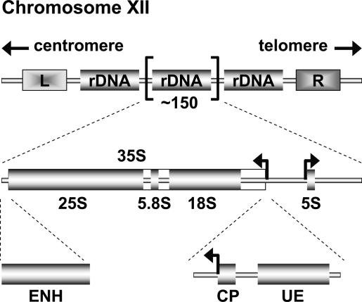
Schematic representation of the yeast rDNA locus. The position of the rDNA repeat cluster on chromosome XII including the left (L) and the right (R) flanking regions with respect to the centromere and telomere is shown. Each rDNA repeat consists of the Pol I-transcribed 35S rRNA gene (precursor for the 18S, 5.8S, and 25S rRNAs) and the RNA Pol III-transcribed 5S rRNA gene. Arrows mark the transcription start sites and direction. The positions of three regulatory DNA elements—ENH, CP, and UE—are indicated.
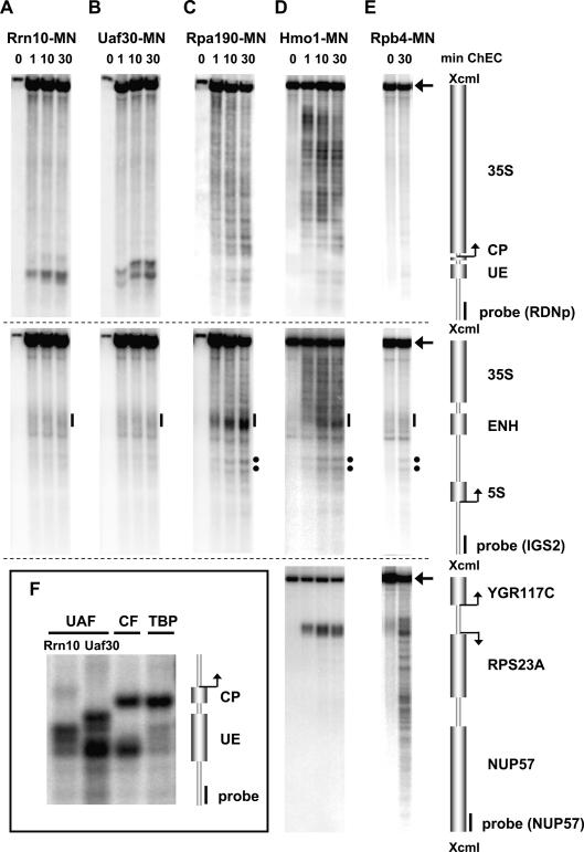
ChEC analyses suggest DNA looping at the yeast rDNA promoter. ChEC was performed with nuclei isolated from yeast strains expressing the MNase fusion protein indicated at the top of each panel. Isolated nuclei were incubated in calcium-containing buffer at 30°C (24°C for Rpb4-MN) for the times depicted above each lane (A–E), or for 30 min (F). (A–F) DNA was isolated, digested with XcmI, separated in a 1% agarose gel, and analyzed in a Southern blot by indirect end-labeling with probes visualizing either a 4.9-kb fragment of the promoter and coding region of the 35S rRNA gene (top panels), a 4.3-kb fragment of IGS1 and IGS2 flanking the 5S rRNA gene, and the 3′ end of the 35S rRNA gene (middle panels), or a 4.2-kb fragment encompassing the RPS23A gene locus (bottom panels). Cartoons of the genomic regions including positions and names of the respective genes with their corresponding transcription start sites, and of probes used for indirect end-labeling, are depicted on the right. Arrowheads point to the uncut XcmI fragment of the corresponding genomic DNA region. (Top panels) (CP) Core promoter; (UE) upstream element; (ENH) enhancer of the 35S rDNA. (Middle panels) Black bars indicate cleavage by promoter-bound factors within the ENH region, and black dots show two hypersensitive sites (C–E). (F) An enlarged view of cleavage events within the rDNA promoter region is presented. In the input lanes of A–C (0) only one-tenth of the DNA was applied to the gel.
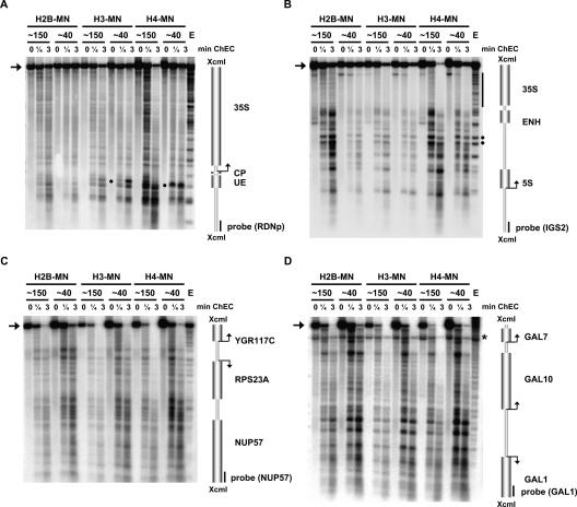
Histone protein association with the 35S rDNA region depends on the transcriptional state of the locus. ChEC was performed with nuclei isolated from yeast strains carrying either a wild-type rDNA locus (∼150) or a locus with a reduced rDNA repeat copy number (∼40) and expressing the MNase fusion protein indicated at the top of the panel. Isolated nuclei were incubated in calcium-containing buffer at 30°C for the minutes depicted above each lane. (Lane E) As a control, nuclei of a wild-type strain were treated with exogeneously added MNase. DNA was analyzed as described in the legend for Figure 2 with probes visualizing either a 4.9-kb fragment of the promoter and coding region of the 35S rRNA gene (A), a 4.3-kb fragment of IGS1 and IGS2 flanking the 5S rRNA gene and the 3′ end of the 35S rRNA gene (B), a 4.2-kb fragment encompassing the RPS23A gene locus (C), or a 6.8-kb fragment containing the GAL gene locus (D). Cartoons of the genomic regions are depicted on the right as described in the legend for Figure 2. Arrowheads on the left point to the uncut XcmI fragment of the corresponding genomic DNA region. (A) Specific cleavage events within the UE occurring selectively in the H3-MNase- and H4-MNase-expressing strains are indicated by black dots on the right. (B) A black bar on the right indicates cleavage within the 3′ region of the 35S rRNA gene, and black dots on the right show two hypersensitive sites at a bidirectional Pol II promoter element (Ganley et al. 2005). (D) An asterisk on the right indicates a background signal from the hybridization with the probe used in B, still detectable after probe removal and rehybridization.
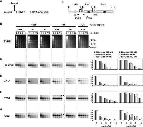
Actively transcribed rRNA genes are largely devoid of histone molecules. (A) Schematic representation of the experimental layout. (B) A cartoon of the yeast rDNA locus is presented. The coding sequences for the ribosomal RNAs (5S, 18S, 5.8S, 25S) and the positions of the two rDNA probes used in the Southern blot analyses (IGS2, ETS1) are indicated. EcoRI restriction sites (E) and positions of the 2.4-kb, 1.9-kb, and 2.8-kb rDNA fragments are depicted. (C–G) ChEC was performed with nuclei isolated from yeast strains carrying either a wild-type rDNA locus (∼150), or a locus with a reduced rDNA repeat copy number (∼40 or ∼25) and expressing the MNase fusion protein indicated at the top of the panel. Before ChEC, purified, linearized plasmid DNA was added to the reaction. Isolated nuclei were incubated in calcium-containing buffer at 30°C for the minutes depicted above each lane. DNA was isolated, digested with EcoRI, and separated in a 1% agarose gel. (C) Analysis by staining with SYBR Safe. Positions and size of three EcoRI fragments originating from the rDNA locus are indicated on the right. (D–G) The DNA was transferred to a nylon membrane and analyzed in a Southern blot with probes indicated on the left, visualizing either a 3.4-kb fragment of plasmid DNA (D), a 1.9-kb fragment containing the GAL gene locus (E), the 1.9-kb fragment containing the 18S rDNA sequence (F), or the 2.4-kb fragment containing the intergenic spacer rDNA region (G). (E,G) Asterisks on the right in lane “∼40 H2B” mark DNA fragments presumably resulting from incomplete EcoRI digestion. Signal intensities of the respective uncut restriction fragments were determined using the FLA-5000 imaging system and MultiGauge software (FujiFilm). The ratios relative to the input value were calculated and plotted against the time of incubation in calcium-containing buffer. A graphical representation of the results is depicted on the right of each row.
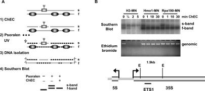
Hmo1 and Histone H3 associate with different states of yeast rDNA chromatin. (A) Schematic representation of a combined ChEC–psoralen cross-linking experiment. An example of specific association of a MNase fusion protein (square) with s-band chromatin is shown. Nucleosomes as components of f-band chromatin are depicted as ovals. Psoralen molecules are represented by black dots, and specific restriction sites used are represented as a pair of scissors. (B) ChEC was performed with nuclei isolated from yeast strains expressing the MNase fusion protein indicated at the top of each panel. Isolated nuclei were incubated in calcium-containing buffer at 30°C for the minutes depicted above each lane. After ChEC, nuclei were treated with psoralen and DNA was isolated. The genomic DNA was either separated in a 1% agarose gel and analyzed by ethidium bromide staining (bottom panel), or digested with EcoRI, separated in a 0.8% agarose gel, and analyzed in a Southern blot with a probe detecting an EcoRI fragment within the coding region of the 35S rRNA gene (top panel). EcoRI restriction sites (E) and the positions of the fragment and probe are indicated in the cartoon below the panels.
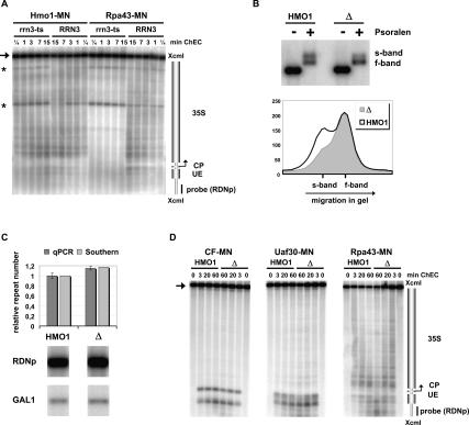
Hmo1 is a structural constituent of actively transcribed 35S rDNA chromatin. (A) Hmo1 remains associated with rDNA in the absence of Pol I. ChEC was performed with nuclei isolated from yeast strains cultured for 2 h at 37°C, carrying either a wild-type (RRN3) or a temperature-sensitive (rrn3-ts) allele of the essential, Pol I-specific initiation factor Rrn3, and expressing the MNase fusion protein indicated at the top of the panel. Isolated nuclei were incubated in calcium-containing buffer at 30°C for the minutes depicted above each lane. DNA was isolated, digested with XcmI, and analyzed in a Southern blot as described in the legend for Figure 2. A cartoon of the genomic region is depicted on the right as described in the legend for Figure 2. An arrow points to the uncut XcmI fragment of the genomic DNA region. Two fragments of unknown origin are labeled by asterisks. (B) S-band chromatin can be established in the absence of Hmo1. Psoralen cross-link analyses were performed with nuclei isolated from yeast strains carrying either a wild-type (HMO1, y881) or a complete deletion (Δ, y1159) of the HMO1 gene. DNA was isolated, digested with EcoRI, and analyzed in a Southern blot as described in the legend for Figure 5B. A profile analysis after quantification of the signal intensity by the FLA-5000 imaging system and MultiGauge software (FujiFilm) is shown. The signal intensity in arbitrary units was plotted against the distance of migration in the gel. (C) rDNA copy number is slightly increased in the HMO1 deletion strain. rDNA copy number was determined in genomic DNA isolated from yeast strains carrying either a wild-type (HMO1, y881) or a complete deletion (Δ, y1159) of the HMO1 gene. The DNA was analyzed by quantitative Southern blot analysis of XcmI restriction fragments detecting either a 4.9-kb fragment of the promoter and coding region of the 35S rRNA gene (RDNp) or a 6.8-kb fragment containing the GAL gene locus (GAL1). Signal intensities of the respective restriction fragments were determined using the FLA-5000 imaging system and MultiGauge software (FujiFilm). The same DNA was analyzed by qPCR amplifying either a region of the 18S rDNA or a region of the single-gene PHO5 locus. The primary data of the Southern blot analysis is shown, with the name of the probes used for hybridization indicated on the left. The diagram is a graphical representation of the results derived by the two kinds of analyses. The relative rDNA copy number was determined by normalizing the ratio of the amount of rDNA to the ratio of the amount of the single-copy genes. (D) Deletion of Hmo1 does not affect transcription factor binding at rDNA. ChEC was performed with nuclei isolated from yeast strains carrying either a wild-type (HMO1) or a complete deletion (Δ) of the HMO1 gene and expressing the MNase fusion protein indicated at the top of the panel. Incubation in calcium-containing buffer was performed for the minutes depicted above each lane. DNA was analyzed as described in the legend for Figure 2. A cartoon of the genomic region is depicted on the right as described in the legend for Figure 2. An arrow points to the uncut XcmI fragment of the genomic DNA region.
References
-
- Bell S.P., Learned R.M., Jantzen H.M., Tjian R. Functional cooperativity between transcription factors UBF1 and SL1 mediates human ribosomal RNA synthesis. Science. 1988;241:1192–1197. - PubMed
-
- Bell S.P., Jantzen H.M., Tjian R. Assembly of alternative multiprotein complexes directs rRNA promoter selectivity. Genes & Dev. 1990;4:943–954. - PubMed
-
- Bier M., Fath S., Tschochner H. The composition of the RNA polymerase I transcription machinery switches from initiation to elongation mode. FEBS Lett. 2004;564:41–46. - PubMed
Publication types
MeSH terms
Substances
LinkOut - more resources
Full Text Sources
Other Literature Sources
Molecular Biology Databases