. 2008 May 1;22(9):1221-30.
doi: 10.1101/gad.1634008.

Cell cycle control of telomere protection and NHEJ revealed by a ts mutation in the DNA-binding domain of TRF2

Affiliations

Cell cycle control of telomere protection and NHEJ revealed by a ts mutation in the DNA-binding domain of TRF2

Akimitsu Konishi et al. Genes Dev. .

Abstract

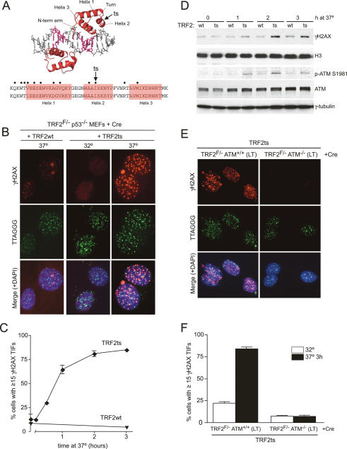
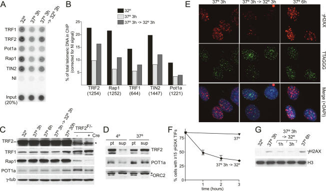
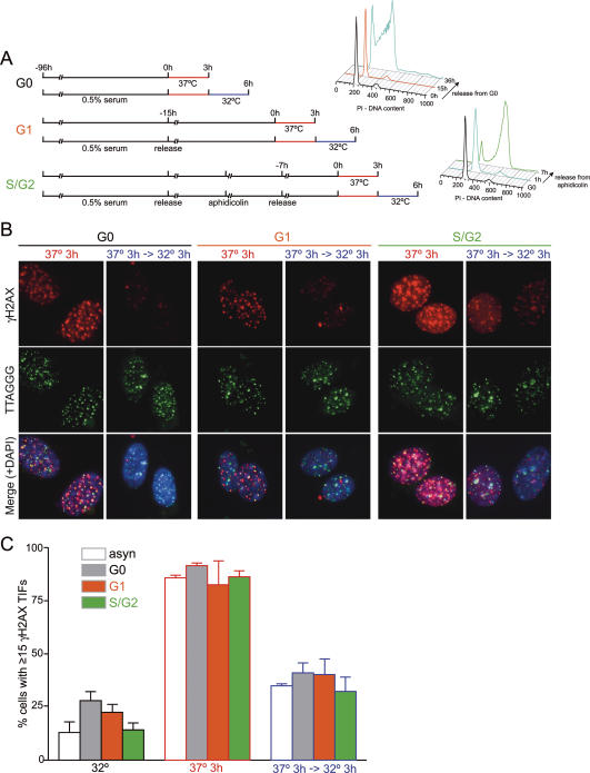
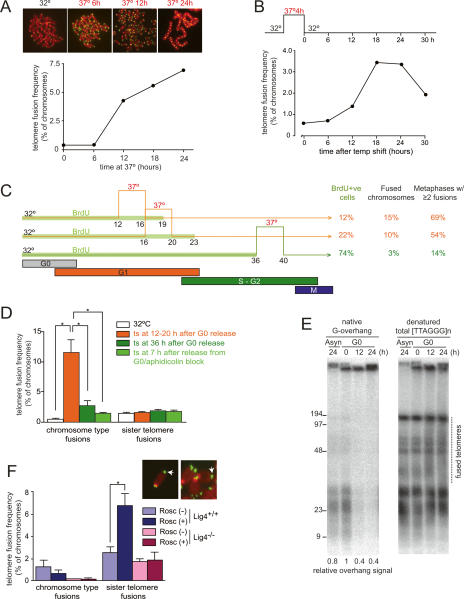
TRF2 is a component of shelterin, the telomere-specific protein complex that prevents DNA damage signaling and inappropriate repair at the natural ends of mammalian chromosomes. We describe a temperature-sensitive (ts) mutation in the Myb/SANT DNA-binding domain of TRF2 that allows controlled and reversible telomere deprotection. At 32 degrees C, TRF2ts was functional and rescued the lethality of TRF2 deletion from conditional TRF2(F/-) mouse embryonic fibroblasts (MEFs). When shifted to the nonpermissive temperature (37 degrees C), TRF2ts cells showed extensive telomere damage resulting in activation of the ATM kinase and nonhomologous end-joining (NHEJ) of chromosome ends. The inactivation of TRF2ts at 37 degrees C was rapid and reversible, permitting induction of short periods (3-6 h) of telomere dysfunction in the G0, G1, and S/G2 phases of the cell cycle. The results indicate that both the induction of telomere dysfunction and the re-establishment of the protected state can take place throughout interphase. In contrast, the processing of dysfunctional telomeres by NHEJ occurred primarily in G1, being repressed in S/G2 in a cyclin-dependent kinase (CDK)-dependent manner.


Figures

Figure 1.
Figure 1.
Generation of a ts allele of TRF2. (A) Schematic of the structure and sequence of the Myb/SANT domain of human and mouse TRF2 highlighting the position of the mutations tested for ts features (see Supplemental Table 1). (Top amino acid sequence) Human amino acids. (Bottom amino acid sequence) Mouse amino acids. The I473A ts mutation (I468A in mouse) is indicated with an arrow. The structure of the TRF2 Myb domains is taken from Rhodes (2005). (B) Temperature-dependent protection of telomeres by TRF2ts. TRF2ts and TRF2wt alleles were expressed in TRF2F/− p53−/− MEFs followed by Cre treatment. Cells were incubated for 3 h at 32°C or 37°C and processed for IF-FISH (γH2AX [red] costained with telomeric TTAGGG-specific FISH probe [green]). The merged images include DAPI staining of DNA (blue). (C) Time course of telomere deprotection at the nonpermissive temperature. TRF2ts and TRF2wt cells were incubated for the indicated time at 37°C, processed for IF-FISH, and scored for 15 or more telomeric γH2AX foci in three independent experiments. Bars indicate standard deviations (SDs). (D) Detection of γH2AX and phosphorylated ATM-S1981. TRF2ts and wild-type TRF2 cells were incubated for the indicated times at 37°C and processed for Western blots. Histone H3, ATM, and γ-tubulin were used as a loading control. (E) ATM dependency of the telomeric DNA damage signal. TRF2ts was expressed in ATM-proficient or -deficient TRF2F/− cells immortalized with SV40-large T (LT) as indicated. TRF2 was deleted with Cre and the cells were processed as in B after 3 h at 37°C. (F) Quantitation of the TIF response in E. Average TIF response values and SDs were derived from three independent experiments.
Figure 2.
Figure 2.
TRF2ts affords transient telomere deprotection through reversible telomere evacuation. (A) ChIP with shelterin components in TRF2ts cells incubated for 3 h at 32°C or 37°C, and for 3 h at 37°C followed by 3 h at 32°C incubation. Crude immune sera were used (serum number, indicated in B). (NI) Nonimmune serum. (B) Quantitative representation of the data in A. Percentage of telomeric DNA for each immunoprecipitation was calculated based on the signal relative to the corresponding total DNA signal. (C) Immunoblot for TRF2 and other shelterin components during short or prolonged incubation at 37°C. TRF2F/−p53−/− MEFs with or without Cre treatment are shown as a control for the endogenous protein levels. (*) Nonspecific signal. (D) Release of TRF2ts and POT1a from nuclei incubated at 37°C. Nuclei from TRF2ts cells were incubated for 30 min at 4°C or 37°C and subjected to centrifugation to separate released proteins (sup) from the nuclei (pt). ORC2 represents a chromatin-bound control. (E) Dissipation of γH2AX TIFs from TRF2ts cells upon shift to 32°C. TRF2ts cells were incubated at 37° and 32°C as indicated and processed to detect TIFs by IF-FISH as described in Figure 1B. (F) Time course of reduction in TIF response after shift to 32°C. TRF2ts cells were shifted for 3 h to 37°C and then incubated for the indicated time periods at either 37°C or 32°C and analyzed by IF-FISH for γH2AX TIFs. The graph shows average values of three experiments and SDs (bars). (G) Reversible induction of H2AX phosphorylation. TRF2ts cells were treated as indicated and analyzed for phosphorylation of H2AX by immunoblotting. Histone H3 serves as a loading control.
Figure 3.
Figure 3.
Loss and re-establishment of telomere protection in G0, G1, and S/G2. (A) Experimental time line for synchronization of TRF2ts cells in G0, G1, and S/G2. TRF2ts cells were arrested in G0 with low serum for 4 d (top) and released into normal medium, and cells in G1 were isolated after 15 h (middle, see FACS profile at 15 h). (Bottom) For S/G2, G0 cells were released into normal medium followed by an aphidicolin block. At 7 h after release from aphidicolin, cells were in S/G2 (see FACS profile). (B) Reversible induction of γH2AX TIFs in G0, G1, and S/G2. TRF2ts cell in G0, G1, S/G2 were incubated for 3 h at 37°C with or without subsequent incubation for 3 h at 32°C and processed for γH2AX TIFs by IF-FISH as in Figure 1B. (C) Quantification of the TIF response. Cells shown in B were scored for 15 or more telomeric γH2AX foci. Bars show average values of three independent experiments and SDs. (Asyn) Asynchronous culture.
Figure 4.
Figure 4.
Cell cycle regulation of telomere fusions. (A) Metaphase spreads with telomeres detected by FISH of TRF2ts cells incubated for the indicated time periods at 37°C (top) and quantitative analysis of telomere fusions (bottom). One-thousand to 1500 chromosomes were scored for fusions. (B) Time course of the occurrence of telomere fusions after incubation at 37°C. TRF2ts cells were incubated for 4 h at 37°C, shifted to 32°C, and incubated for the indicated time. Telomere fusions were scored on 1000–1500 metaphase chromosomes per time point. (C) Incidence of telomere fusions after inactivation of TRF2ts in G0, G1, and S/G2. TRF2ts cells were arrested in G0 by serum starvation and released into serum-containing medium containing BrdU. Cells were incubated for 4 h at 37°C at 12 and 16 h after release when cells are in G1, and at 36 h when cells are in S/G2. Cells were shifted back to 32°C and harvested at 45 h after G0 release. Prior to preparation of metaphase spreads, cells were incubated for 2 h with colcemid. BrdU incorporation was determined by FACS. (D) Quantification of telomere fusions using the experimental setup shown in C. Bars represent mean values from three experiments and SDs (error bar). (*) P < 0.05, based on nonpaired Student’s t-test. (E) NHEJ processing of telomeres in G0. DNA was isolated from TRF2ts cells treated for the times indicated above the lanes at 37°C ([Asyn] asynchronous population) and analyzed by in-gel hybridization for the status of the telomeric overhang and telomere fusions. Molecular weights are indicated in kilobases. (F) Inhibition of CDK activity with roscovitine induces sister telomere fusions in S/G2. TRF2ts cells with or without DNA ligase IV were arrested in G0, released into normal medium followed by aphidicolin block, and released again to proceed into S/G2 for 7 h. Cells were incubated for 4 h at 37°C with or without roscovitine and telomere fusions were scored as in D. (Inset) Metaphase chromosomes with sister fusions (arrowheads).

Similar articles

Cited by

References

    1. Aylon Y., Kupiec M. Cell cycle-dependent regulation of double-strand break repair: A role for the CDK. Cell Cycle. 2005;4:259–261. - PubMed
    1. Aylon Y., Liefshitz B., Kupiec M. The CDK regulates repair of double-strand breaks by homologous recombination during the cell cycle. EMBO J. 2004;23:4868–4875. - PMC - PubMed
    1. Bailey S.M., Cornforth M.N., Kurimasa A., Chen D.J., Goodwin E.H. Strand-specific postreplicative processing of mammalian telomeres. Science. 2001;293:2462–2465. - PubMed
    1. Bilaud T., Brun C., Ancelin K., Koering C.E., Laroche T., Gilson E. Telomeric localization of TRF2, a novel human telobox protein. Nat. Genet. 1997;17:236–239. - PubMed
    1. Broccoli D., Smogorzewska A., Chong L., de Lange T. Human telomeres contain two distinct Myb-related proteins, TRF1 and TRF2. Nat. Genet. 1997;17:231–235. - PubMed

Publication types

MeSH terms

Substances