. 2008 May 1;22(9):1231-43.
doi: 10.1101/gad.1643308.

Fibronectin fibrillogenesis regulates three-dimensional neovessel formation

Affiliations

Fibronectin fibrillogenesis regulates three-dimensional neovessel formation

Xiaoming Zhou et al. Genes Dev. .

Abstract

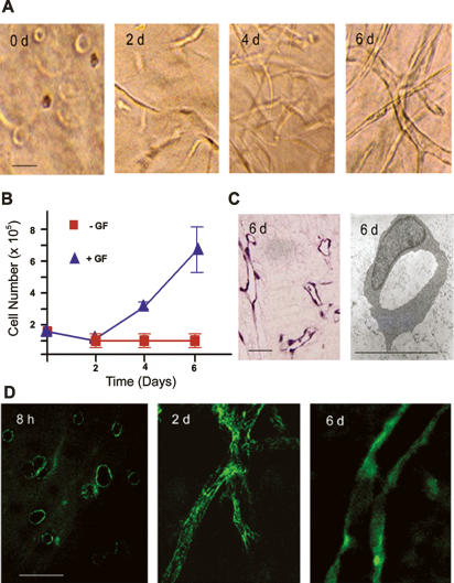
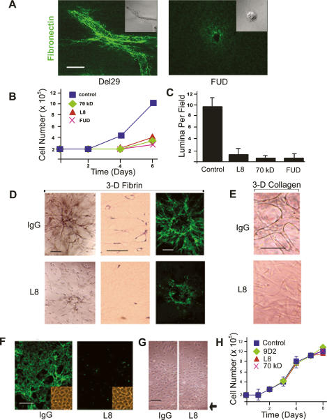
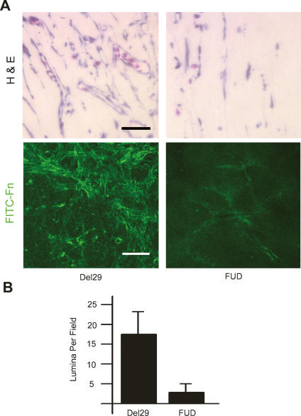
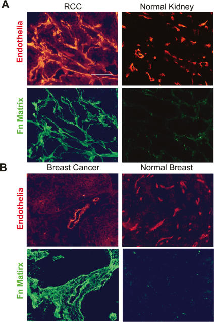
During vasculogenesis and angiogenesis, endothelial cell responses to growth factors are modulated by the compositional and mechanical properties of a surrounding three-dimensional (3D) extracellular matrix (ECM) that is dominated by either cross-linked fibrin or type I collagen. While 3D-embedded endothelial cells establish adhesive interactions with surrounding ligands to optimally respond to soluble or matrix-bound agonists, the manner in which a randomly ordered ECM with diverse physico-mechanical properties is remodeled to support blood vessel formation has remained undefined. Herein, we demonstrate that endothelial cells initiate neovascularization by unfolding soluble fibronectin (Fn) and depositing a pericellular network of fibrils that serve to support cytoskeletal organization, actomyosin-dependent tension, and the viscoelastic properties of the embedded cells in a 3D-specific fashion. These results advance a new model wherein Fn polymerization serves as a structural scaffolding that displays adhesive ligands on a mechanically ideal substratum for promoting neovessel development.


Figures

Figure 1.
Figure 1.
Endothelial cell tubulogenesis and Fn matrix assembly. (A) Endothelial cells (1.5 × 10⁵) were embedded in a 3D fibrin gel in the presence of 20% human serum and a cocktail of VEGF, HGF, TGFα, TGFβ1, and heparin. Phase contrast micrographs are shown following 0, 2, 4, and 6 d in culture. (B) Endothelial cell growth in the presence or absence of the provasculogenic cocktail was assessed by protease digestion of the 3D fibrin matrix followed by counting of cells. (C) Following 6 d in culture, patent endothelial cell tubules are formed as assessed in H&E-stained cross-sections or TEMs. (D) Confocal laser micrographs of pericellular assembly of FITC-labeled Fn matrix following 8 h, 2 d, and 6 d of culture. Bar, 30 μm.
Figure 2.
Figure 2.
Fn matrix assembly regulates endothelial cell 3D morphogenesis. (A) Endothelial cells were suspended in 3D fibrin gels with FITC-labeled Fn in the absence or presence of the FUD peptide (250 nM), with matrix assembly and cell morphology monitored by confocal laser microscopy. Bar, 20 μm. (B) Endothelial cells were cultured within 3D fibrin matrices for 6 d in the presence or absence of Fn matrix inhibitors. Cell number was quantified following gel digestion with collagenase. (C) Endothelial cells were cultured in 3D fibrin gels under control conditions or in the presence of various Fn matrix assembly inhibitors for 6 d, and tubulogenesis was assessed by sectioning and H&E staining of the matrices followed by lumen quantification. (D) Endothelial cells spheroids were embedded in gels of 3D fibrin and cultured for 6 d in the absence or presence of mAb L8 (100 μg/uL), and tubulogenesis was assessed by phase contrast microscopy (left panels in the top and bottom series) or light microscopy (H&E-stained cross-sections in the middle column). (Right panels) The assembly of a FITC-labeled Fn matrix was monitored by confocal laser microscopy. Bar, 50 μm. (E) Endothelial cells were cultured within a matrix of type I collagen in the presence or absence of mAb L8 and tubulogenesis assessed at 6 d by phase contrast microscopy. Bar, 50 μm. (F) Endothelial cells were cultured atop 3D fibrin gels for 6 d in the presence of L8 or control IgG. (Inset) Assembly of FITC-Fn into a fibrillar matrix was monitored by fluorescence microscopy, and cell morphology was assessed by phase contrast microscopy. (G) Endothelial cell 2D migration was assessed atop a fibrin-coated substratum in the presence of IgG or L8 (arrowhead marks the edge of the monolayer at the start of the 2-d culture period). (H) Endothelial cell growth was monitored during a 6-d culture period in the presence of mAb L8, mAb 9D2, or the 70-kDa Fn fragment relative to an IgG control. Bar, 100 μm.
Figure 3.
Figure 3.
Fn matrix assembly is required for angiogenesis in vivo. (A) Type I collagen/fibrin composite gels polymerized in perforated transwell chambers were prepared. A Matrigel reservoir containing 200 ng of VEGF and 100 ng of HGF with either the Del29 control peptide or the FUD peptide was placed atop the matrix. (Top panels) The apparatus was placed atop the dropped CAM, and angiogenesis was allowed to proceed. After 3 d, the gels were harvested and vascular ingrowth was monitored by light microscopy following H&E staining (bar, 40 μm). (Bottom panels) In some experiments, FITC-Fn was supplemented in the matrices during the culture period, and Fn fibrillogenesis within the gels was monitored by confocal laser microscopy (bar, 50 μm). (B) After sectioning, lumina per 40× field were counted.
Figure 4.
Figure 4.
Fn matrix assembly is restricted to human neovessels in vivo. (A) Renal cell carcinoma (left panels) or normal kidney (right panels; n = 8) were stained for UEA-1 (top panels, red) or with mAb L8 (bottom panels, green). (B) Breast carcinoma (left panels) was stained for UEA-1 (top left panel, red), or with mAb L8 (bottom left panel, green). Normal breast (n = 8) was stained for FVIIIRAg (top right panel, red), or with mAb L8 (bottom right panel, green).
Figure 5.
Figure 5.
The 3D Fn matrix is required for endothelial cell cytoskeletal organization and adhesion. (A) Levels of phosphorylated ERK1/2, JNK, and p38 were determined by immunoblot analysis in lysates of endothelial cells embedded in fibrin gels in the presence of either control IgG or mAb L8 for 0 h, 2 h, 1 d, or 2 d. Total ERK1/2 serves as the loading control. (B) Endothelial cells were cultured in 3D fibrin gels in the presence of the FUD or control peptides for 2 d. F-actin cytoskeletal organization was monitored following staining with Alexa 488-conjugated phalloidin and confocal laser microscopy. Bar, 20 μm. (C) Endothelial cells in 3D fibrin matrices in the presence or absence of FUD were either stained with an antibody against activated β1 integrin (green, left panels) or transfected with a GFP-tagged vinculin expression vector (pRK-vinculin-EGFP, green, right panels). Following counterstaining with Alexa 594-labeled phalloidin (red), fluorescence was monitored by confocal laser microscopy. Bar, 10 μm. (D) Endothelial cells were cultured atop a 2D fibrin gel for 2 d in the presence of either Del29 or FUD peptides. In tandem with staining with Alexa 594-labeled phalloidin to monitor cytoskeletal organization, actve β1 integrin and vinculin distribution were monitored by staining with an antibody against active β1 integrin (left panels) or assessing GFP-vinculin localization (right panels), respectively, by confocal microscopy. Bar, 20 μm.
Figure 6.
Figure 6.
Fn matrix assembly regulates the generation of 3D-specific myosin-dependent tension and intracellular rigidity. (A) 3D fibrin gels were cast in individual wells of 24-well plates and cultured alone or in the presence of embedded endothelial cells for 2 d in the presence of control IgG, mAb L8, mAb 9D2, the 70-kDa Fn fragment, or the FUD peptide. Gels were detached from the edges of the culture wells and contraction-monitored after an additional incubation period of 10 h at 37°C. The percentage inhibition of gel contraction (measured as the change in gel diameter) relative to an IgG control is presented. Inset shows photographs (from left to right) of a cell-free fibrin gel, a gel contracted by embedded endothelial cells, and gels contracted by endothelial cells cultured in the presence of either mAb 9D2 or mAb L8 or the 70-kDa Fn fragment. (B) Endothelial cells were cultured in 3D fibrin gels for 2 d with either the FUD peptide or the control Del29 peptide. Levels of β-actin, α-actinin, NMMIIA, NMMIIB, and MLC2 were measured by Western blot, with ERK1/2 serving as the loading control. As assessed by semiquantitative densitometry, the levels of β-actin, actinin, and MLC2 were 58 ± 7% (n = 5; mean ± 1 SD), 62% (n = 2), and 60 ± 12% (n = 3; mean ± 1 SD) of control. (C) Prior to embedding in the 3D fibrin matrix, endothelial cells were ballistically microinjected with 100-nm polystyrene beads. After 3-d incubation, the beads dispersed in the cytoplasm and their centroids were tracked with high spatial and temporal resolution using fluorescence microscopy. Bar, 10 μm. (D) Typical trajectories of beads in the cytoplasm of 3D-embedded cells, under control conditions (black, left panel) and in the presence of FUD (green, right panel). Time of movie capture, 20 sec. Bar, 50 nm. (E) Ensemble-averaged MSDs of beads in the cytoplasm of 3D-embedded endothelial cells under control conditions (black curve) and in the presence of FUD (green curve) or blebbistatin (blue curve). MSDs were computed from displacements of the beads such as shown in D. At least 110 beads in at least 10 cells were tracked for each condition. (F) Averaged elastic modulus of cells under control conditions or in the presence of FUD or blebbistatin. (***) P < 0.001. (G) Endothelial cells were cultured in 3D fibrin gels for 2 d in the presence or absence of 50 μM ± blebbistatin when F-actin organization and Fn matrix assembly were monitored by confocal laser microscopy. (Top panels) F-actin organization was monitored by staining with Alexa 488-conjugated phalloidin. (Bottom panels) Fn matrix assembly was assessed by culture in the presence of FITC-conjugated Fn. Bar, 20 μm. (H) Endothelial cells were cultured in 3D fibrin gels for 6 d under provasculogenic conditions in the presence of 50 μM ± blebbistatin or vehicle (DMSO). Tubulogenesis was monitored following sectioning, H&E staining, and lumen quantification.

Similar articles

Cited by

References

    1. Ambesi A., Klein R.M., Pumiglia K.M., McKeown-Longo P.J. Anastellin, a fragment of the first type III repeat of fibronectin, inhibits extracellular signal-regulated kinase and causes G(1) arrest in human microvessel endothelial cells. Cancer Res. 2005;65:148–156. - PubMed
    1. Baneyx G., Baugh L., Vogel V. Fibronectin extension and unfolding within cell matrix fibrils controlled by cytoskeletal tension. Proc. Natl. Acad. Sci. 2002;99:5139–5143. - PMC - PubMed
    1. Beningo K.A., Dembo M., Wang Y.L. Responses of fibroblasts to anchorage of dorsal extracellular matrix receptors. Proc. Natl. Acad. Sci. 2004;101:18024–18029. - PMC - PubMed
    1. Ben-Ze’ev A., Farmer S.R., Penman S. Protein synthesis requires cell-surface contact while nuclear events respond to cell shape in anchorage-dependent fibroblasts. Cell. 1980;21:365–372. - PubMed
    1. Bershadsky A., Kozlov M., Geiger B. Adhesion-mediated mechanosensitivity: A time to experiment, and a time to theorize. Curr. Opin. Cell Biol. 2006;18:472–481. - PubMed

Publication types

MeSH terms

LinkOut - more resources