. 2008 May 1;22(9):1244-56.
doi: 10.1101/gad.1632308.

PX-RICS mediates ER-to-Golgi transport of the N-cadherin/beta-catenin complex

Affiliations

PX-RICS mediates ER-to-Golgi transport of the N-cadherin/beta-catenin complex

Tsutomu Nakamura et al. Genes Dev. .

Abstract

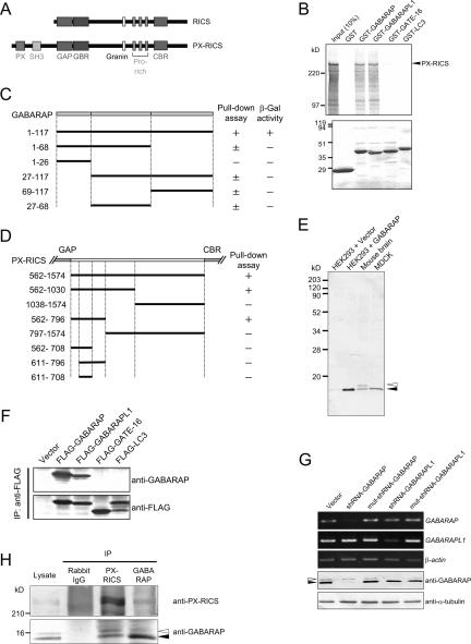
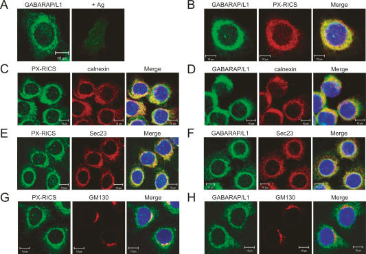
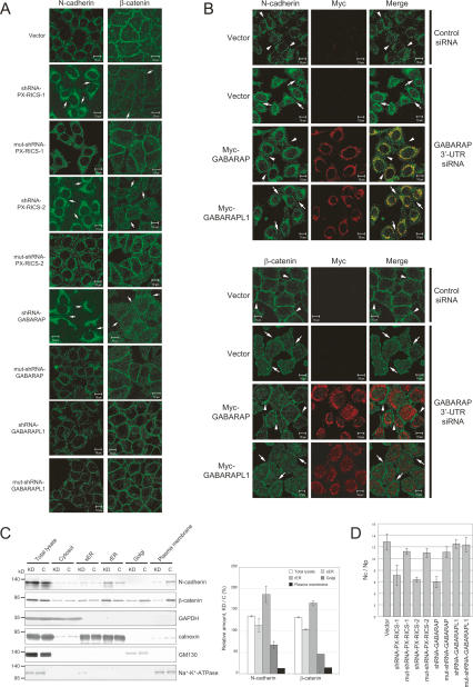
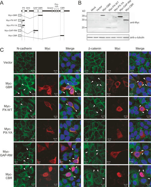
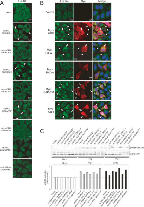
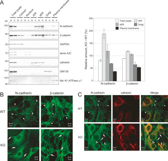
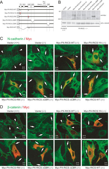
Cadherins mediate Ca2+-dependent cell-cell adhesion. Efficient export of cadherins from the endoplasmic reticulum (ER) is known to require complex formation with beta-catenin. However, the molecular mechanisms underlying this requirement remain elusive. Here we show that PX-RICS, a beta-catenin-interacting GTPase-activating protein (GAP) for Cdc42, mediates ER-to-Golgi transport of the N-cadherin/beta-catenin complex. Knockdown of PX-RICS expression induced the accumulation of the N-cadherin/beta-catenin complex in the ER and ER exit site, resulting in a decrease in cell-cell adhesion. PX-RICS was also required for ER-to-Golgi transport of the fibroblast growth factor-receptor 4 (FGFR4) associated with N-cadherin. PX-RICS-mediated ER-to-Golgi transport was dependent on its interaction with beta-catenin, phosphatidylinositol-4-phosphate (PI4P), Cdc42, and its novel binding partner gamma-aminobutyric acid type A receptor-associated protein (GABARAP). These results suggest that PX-RICS ensures the efficient entry of the N-cadherin/beta-catenin complex into the secretory pathway, and thereby regulates the amount of N-cadherin available for cell adhesion and FGFR4-mediated signaling.


Figures

Figure 1.
Figure 1.
PX-RICS interacts with GABARAP/L1 in vitro and in vivo. (A) Schematic representation of RICS and PX-RICS. The domains and motifs contained in RICS and PX-RICS are shown. (PX) Phox homology domain; (SH3) Src homology 3 domain; (GAP) GTPase-activating protein domain; (GBR,) GABARAP-binding region; (Granin) granin motif; (Pro-rich) polyproline stretch; (CBR) β-catenin-binding region. (B) Association of GABARAP and GABARAPL1 with PX-RICS in vitro. In vitro translated 35S-labeled PX-RICS was incubated with GST or GST fusion proteins as indicated and bound proteins were analyzed. The bottom panel shows Coomassie staining of GST and GST fusion proteins used in pull-down assays. (C) Mapping of regions in GABARAP required for binding to PX-RICS. GABARAP was divided into three regions based on the ability of each to interact with other known partners, including GABAARγ2, tubulin, gephyrin, NSF, and Unc-51-like kinase 1 (ULK1). GABARAP fragments fused to GST were analyzed for their ability to interact with in vitro translated 35S-labeled PX-RICS. The results of yeast two-hybrid assays are also shown. (+) Detectable activity; (±) residual activity; (−) no detectable activity. (D) Mapping of regions in PX-RICS required for binding to GABARAP. In vitro translated 35S-labeled fragments of PX-RICS were analyzed for their ability to interact with GST-GABARAP. (+) Detectable activity; (−) no detectable activity. (E) Detection of endogenous GABARAP/L1 with anti-GABARAP antibody. Lysates prepared from HEK293 cells transfected with empty vector or GABARAP, mouse brain, or MDCK cells were immunoblotted with anti-GABARAP antibody. The 15- and 16-kDa proteins are indicated by the solid and open arrowheads, respectively. (F) Specificity of anti-GABARAP antibody. HEK293 cells transfected with Flagtagged GABARAP, GABARAPL1, GATE-16, orMAP1A/1B LC3 were subjected to immunoprecipitation with anti-Flag antibody followed by immunoblotting with anti-GABARAP or anti-Flag antibody. (G) The 15- and 16-kDa proteins are down-regulated specifically by shRNAs against GABARAP and GABARAPL1, respectively. HeLa cells were transfected with empty vector, shRNA-GABARAP, shRNA-GABARAPL1, or their mutant forms, mut-shRNA-GABARAP or mut-shRNA-GABARAPL1, and subjected to semiquantitative RT–PCR. Cell lysates were subjected to immunoblotting analysis with anti-GABARAP antibody. GABARAP and GABARAPL1 are indicated by the solid and open arrowheads, respectively. Anti-α-tubulin antibody was used as a control. (H) Association of GABARAP/L1 with PX-RICS in vivo. HeLa cell lysates were subjected to immunoprecipitation, followed by immunoblotting with the indicated antibodies. GABARAP and GABARAPL1 are indicated by the solid and open arrowheads, respectively.
Figure 2.
Figure 2.
PX-RICS and GABARAP/L1 are colocalized at the ER and ER exit site. (A) HeLa cells were immunostained with anti-GABARAP antibody. (+Ag) Anti-GABARAP antibody was preabsorbed with immunizing antigen prior to use. (B) Colocalization of PX-RICS and GABARAP/L1 at the perinuclear region. HeLa cells were double-labeled with anti-PX-RICS and anti-GABARAP antibodies. (C–H) Subcellular localization of PX-RICS and GABARAP/L1. HeLa cells were double-labeled with the indicated antibodies. Bars, 10 μm.
Figure 3.
Figure 3.
PX-RICS and GABARAP are critical for ER-to-Golgi transport of the N-cadherin/β-catenin complex. (A) Knockdown of PX-RICS or GABARAP results in the disappearance of N-cadherin and β-catenin at the cell–cell boundaries and intracellular accumulation of N-cadherin. HeLa cells were transfected with the indicated shRNAs and subjected to immunofluorescent staining with anti-N-cadherin or anti-β-catenin antibody. Arrows indicate the absence of N-cadherin and β-catenin at the borders between two neighboring cells. Bars, 10 μm. (B) Exogenous expression of GABARAP, but not GABARAPL1, restores the subcellular distribution of N-cadherin and β-catenin in GABARAP-knockdown cells. HeLa cells were cotransfected with siRNA for GABARAP 3′-UTR and Myc-tagged GABARAP or GABARAPL1, and subjected to immunostaining with the indicated antibodies. Arrowheads indicate N-cadherin or β-catenin enriched at the sites of cell–cell contact. The cell–cell boundaries lacking N-cadherin or β-catenin are indicated by arrows. Bars, 10 μm. (C) Subcellular distribution of N-cadherin and β-catenin in PX-RICS-knockdown (KD) and control (C) HeLa cells. (Left panel) After subcellular fractionation, the amounts of N-cadherin and β-catenin in each fraction were evaluated by immunoblotting. (Right panel) The results were analyzed by densitometry and the relative amounts in knockout versus control cells were quantified (mean ± SD [n = 3]). Each fraction was analyzed for the distribution of marker proteins specific for various organelles: GAPDH (cytosol), calnexin (ER), GM130 (Golgi), and Na+/K+-ATPase α1 (plasma membrane). (D) Ca2+-dependent cell adhesion is abrogated by knockdown of PX-RICS or GABARAP. HeLa cells expressing the indicated shRNA were dissociated by pipetting in the presence of Ca2+. Cell adhesion activity was quantified by counting the total number of cells (Nc) and the number of particles (cell clumps) (Np). Error bars represent the mean ± SD (n = 5).
Figure 4.
Figure 4.
ER-to-Golgi transport of the N-cadherin/β-catenin complex is dependent on the interaction of PX-RICS with its associated molecules. (A) Schematic representation of the dominant-negative mutants of PX-RICS that block the interaction of PX-RICS with its associated molecules. (PX) Phox homology domain; (SH3) Src homology 3 domain; (GAP) GTPase-activating protein domain; (GBR) GABARAP-binding region; (Granin) granin motif; (Pro-rich) polyproline stretch; (CBR) β-catenin-binding region. Shaded rectangles indicate Myc tag. (B) Protein expression of dominant-negative mutants of PX-RICS in HeLa cells. Lysates prepared from HeLa cells expressing the indicated Myc-tagged fragments of PX-RICS were immunoblotted with anti-Myc tag antibody. Anti-α-tubulin antibody was used as a control. (C) Block of the interaction between PX-RICS and its associated molecules results in the disappearance of N-cadherin and β-catenin from the cell–cell boundaries. HeLa cells were transfected with the indicated Myc-tagged fragments of PX-RICS and processed for immunostaining with anti-Myc tag antibody plus anti-N-cadherin or anti-β-catenin antibody. N-cadherin and β-catenin disappeared from the cell–cell boundaries of HeLa cells expressing dominant-negative mutants of PX-RICS (arrowheads). Bars, 10 μm.
Figure 5.
Figure 5.
The N-cadherin/β-catenin complex is accompanied by FGFR4 in PX-RICS/GABARAP-mediated transport. (A) Knockdown of PX-RICS or GABARAP induces the accumulation of FGFR4 in the perinuclear ER-like compartments. HeLa cells were transfected with shRNAs or mutant shRNAs as indicated and probed with anti-FGFR4 antibody. Perinuclear accumulation of FGFR4 is indicated by arrows. Bars, 10 μm. (B) Accumulation of FGFR4 in HeLa cells expressing dominant-negative mutants of PX-RICS. HeLa cells were transfected with Myc-tagged fragments of PX-RICS and processed for immunostaining with anti-FGFR4 and anti-Myc tag antibodies. Note that FGFR4 is accumulated in Myc-positive cells (arrows), including those with low expression levels. Arrowheads indicate the surrounding untransfected cells. Bars, 10 μm. (C) FGF signaling is suppressed by knockdown of PX-RICS or GABARAP. HeLa cells expressing the indicated shRNAs were assessed for FGF1- or FGF2-induced activation of p42/44 MAPK, downstream effecters of FGF signaling, by immunoblotting with antibodies specific for the phosphorylated (activated) form of p42/44 (phospho-p42/44) and with antibodies against all forms of p42/44 (total p42/44) as indicated. The results were quantified by densitometry and the ratio of phosphorylated to total p42/44 in each sample is shown.
Figure 6.
Figure 6.
PX-RICS regulates ER-to-Golgi transport of N-cadherin and β-catenin in MEFs. (A) Subcellular distribution of N-cadherin and β-catenin in MEFs. (Left panel) Subcellular fractionation was performed for wild-type (W) and PX-RICS-knockout (K) MEFs, and the amounts of N-cadherin and β-catenin in each fraction were evaluated by immunoblotting. (Right panel) The results were analyzed by densitometry and the relative amounts in knockout versus wild-type cells were quantified (mean ± SD [n = 3]). Each fraction was analyzed for the distribution of marker proteins specific for various organelles: GAPDH (cytosol), lamin A/C (nuclei), calnexin (ER), GM130 (Golgi), and Na+/K+-ATPase α1 (plasma membrane). (B) Immunocytochemical localization of N-cadherin and β-catenin in PX-RICS−/− MEFs. Primary MEFs prepared from wild-type (WT) or PX-RICS−/− (KO) mice were probed with antibodies to N-cadherin or β-catenin. Arrowheads indicate N-cadherin or β-catenin enriched at the sites of cell–cell contact. The cell–cell contact sites lacking N-cadherin or β-catenin are indicated by arrows. Bars, 10 μm. (C) ER accumulation of N-cadherin in PX-RICS−/− MEFs. Wild-type (WT) or PX-RICS−/− (KO) MEFs were processed for double-staining with anti-N-cadherin and anti-calnexin antibodies. Arrowheads indicate N-cadherin enriched at the sites of cell–cell contact. The cell–cell contact sites lacking N-cadherin are indicated by arrows. Bars, 10 μm.
Figure 7.
Figure 7.
Exogenous expression of PX-RICS, but not its mutant forms, restores the subcellular distribution of N-cadherin and β-catenin. (A) Schematic representation of the PX-RICS mutants. (PX) Phox homology domain; (SH3) Src homology 3 domain; (GAP) GTPase-activating protein domain; (GBR) GABARAP-binding region; (Granin) granin motif; (Pro-rich) polyproline stretch; (CBR) β-catenin-binding region. Shaded rectangles indicate Myc tag. (B) Protein expression of wild-type and mutant forms of PX-RICS and RICS in MEFs. Wild-type and PX-RICS−/− MEFs were transfected with indicated Myc-tagged expression plasmids, and the cell lysates were immunoblotted with anti-Myc tag antibody. Anti-α-tubulin antibody was used as a control. (C,D) Exogenous expression of PX-RICS, but not its mutant forms, restores the subcellular distribution of N-cadherin (C) and β-catenin (D) in PX-RICS−/− MEFs. Primary PX-RICS−/− MEFs were transfected with Myc-tagged wild-type or mutant PX-RICS, and were subjected to immunostaining with the indicated antibodies. Arrowheads indicate N-cadherin or β-catenin enriched at the sites of cell–cell contact. The cell–cell contact sites lacking N-cadherin or β-catenin are indicated by arrows. Bars, 10 μm.

Similar articles

Cited by

References

    1. Audhya A., Foti M., Emr S.D. Distinct roles for the yeast phosphatidylinositol 4-kinases, Stt4p and Pik1p, in secretion, cell growth, and organelle membrane dynamics. Mol. Biol. Cell. 2000;11:2673–2689. - PMC - PubMed
    1. Barlowe C. Signals for COPII-dependent export from the ER: What’s the ticket out? Trends Cell Biol. 2003;13:295–300. - PubMed
    1. Bole D.G., Hendershot L.M., Kearney J.F. Posttranslational association of immunoglobulin heavy chain binding protein with nascent heavy chains in nonsecreting and secreting hybridomas. J. Cell Biol. 1986;102:1558–1566. - PMC - PubMed
    1. Bruns J.R., Ellis M.A., Jeromin A., Weisz O.A. Multiple roles for phosphatidylinositol 4-kinase in biosynthetic transport in polarized Madin-Darby canine kidney cells. J. Biol. Chem. 2002;277:2012–2018. - PubMed
    1. Cavallaro U., Niedermeyer J., Fuxa M., Christofori G. N-CAM modulates tumour-cell adhesion to matrix by inducing FGF-receptor signalling. Nat. Cell Biol. 2001;3:650–657. - PubMed

Publication types

MeSH terms