. 2008 May 1;68(9):3204-13.
doi: 10.1158/0008-5472.CAN-07-2949.

CD151 accelerates breast cancer by regulating alpha 6 integrin function, signaling, and molecular organization

Affiliations

CD151 accelerates breast cancer by regulating alpha 6 integrin function, signaling, and molecular organization

Xiuwei H Yang et al. Cancer Res. .

Abstract

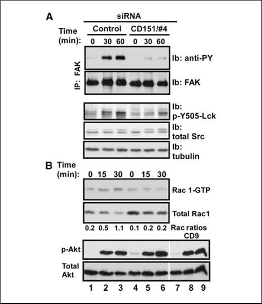
CD151, a master regulator of laminin-binding integrins (alpha(6)beta(4), alpha(6)beta(1), and alpha(3)beta(1)), assembles these integrins into complexes called tetraspanin-enriched microdomains. CD151 protein expression is elevated in 31% of human breast cancers and is even more elevated in high-grade (40%) and estrogen receptor-negative (45%) subtypes. The latter includes triple-negative (estrogen receptor, progesterone receptor, and HER2 negative) basal-like tumors. CD151 ablation markedly reduced basal-like mammary cell migration, invasion, spreading, and signaling (through FAK, Rac1, and lck) while disrupting epidermal growth factor receptor (EGFR)-alpha(6) integrin collaboration. Underlying these defects, CD151 ablation redistributed alpha(6)beta(4) integrins subcellularly and severed molecular links between integrins and tetraspanin-enriched microdomains. In a prototypical basal-like mammary tumor line, CD151 ablation notably delayed tumor progression in ectopic and orthotopic xenograft models. These results (a) establish that CD151-alpha(6) integrin complexes play a functional role in basal-like mammary tumor progression; (b) emphasize that alpha(6) integrins function via CD151 linkage in the context of tetraspanin-enriched microdomains; and (c) point to potential relevance of CD151 as a high-priority therapeutic target, with relative selectivity (compared with laminin-binding integrins) for pathologic rather than normal physiology.


Figures

Figure 1
Figure 1
CD151 protein expression in human breast carcinoma. A, immunohistochemistry for CD151 was done on paraffin sections of tissue microarrays containing samples of normal breast tissue (a and b) and invasive breast tumors (c–h). a, normal breast duct (×40). b, normal breast lobule (×100); arrows, representative CD151-positive cells in the basal/myoepithelial layer. c and d, human tumors with absent and low degree of CD151 immunostaining, respectively (×40). e and f, human tumors with moderate 2+ overexpression of CD151 predominantly located on the membrane or in cytoplasm, respectively (×40). g and h, human tumors with marked 3+ overexpression of CD151 on the membrane or in cytoplasm, respectively (×40). B, human breast cancer tissue microarray samples (a total of 124 patients) were subdivided according to modified Bloom-Richardson grade and estrogen receptor protein expression. The percent of each subgroup expressing high CD151 (3+ or 2+) is indicated. For further details, see Supplementary Table S1.
Figure 2
Figure 2
CD151 supports mammary epithelial cell migration and invasion. A, after treatment with siRNAs, MCF-10A cells, grown to confluence, were then incubated in a 24-well plate with serum-free medium containing 10 μg/mL mitomycin C at room temperature for 1 h. After gaps were scratched into cell monolayers, 0.5 mL of serum-free medium containing 10 ng/mL EGF was added, and gaps were evaluated after 0 and 18 h at 37°C. Percentage of gap closure was determined by measuring the mean change in gap width at three representative sites in three independent experiments (n = 3) *, P < 0.05. Right, siRNA knockdown efficiency. MCF-10A cells in 24-well plates were treated with siRNAs [mock, control (Cntl), or CD151#4] for 5 d, and cell lysates (in RIPA buffer) were blotted for CD151 (mAb 1A5) and β-actin. B, after treatment with siRNAs, MDA-MB-231 cells (5 × 10⁴) in serum-free medium containing 0.1% BSA were added to the top of Matrigel-coated transwell chambers. Serum-free medium (0.75 mL) containing 10 ng/mL EGF and 0.1% BSA was added to the bottom of transwell chambers. After ~18 h at 37°C, cells that had invaded through the Matrigel were fixed, stained, and photographed, and the mean number of invaded cells was determined from triplicate chambers. Bar, 100 μm. Right, siRNA efficiency. MDA-MB-231 cells were treated with siRNAs and then lysates were blotted for tetraspanins CD151 (mAb 1A5) and CD9 (mAb MM2/57). The two rows of numbers below the figure indicate percent knockdown values for CD151 and CD9, respectively, as determined by densitometry. C, after treatment with siRNAs (alone or in combination), MDA-MB-231 cells were again analyzed for invasion through Matrigel, as in B. D, a metastatic mouse mammary tumor cell line (J110) was treated with siRNA to murine CD151 (70% knockdown) or murine α6 integrin (>80% knockdown). Invasion was then analyzed as in B. *, P < 0.05; **, P < 0.01.
Figure 3
Figure 3
CD151 effects on EGF-stimulated mammary cell spreading and invasion. A, after siRNA treatment, MDA-MB-231 cells were suspended in serum-free medium at 37°C for 45 min, and then plated onto laminin-1–or fibronectin (Fn)-coated 24-well plates with or without EGF (10 ng/mL). After 45 min, representative fields were photographed. B, percentages of spread cells were determined (n = 4). Note: No cell spreading was observed on plastic surfaces coated with BSA (data not shown). C, invasion was also analyzed (as in Fig. 2B) with or without EGF (10 μg/mL) added to the bottom of the invasion chamber. After tumor formation in nude mice, MDA-MB-231 cells were reisolated (now called p-MDA-MB-231). These passaged sublines were isolated from tumors originating from MDA-MB-231 cells that had been treated with control shRNA (C1) or CD151 shRNA (K1 and K2; see also Fig. 6C). D, a subline of MCF-7 was enriched for Hoechst dye exclusion and elevated EGFR (now called s-MCF-7) and was analyzed for invasion as in C.
Figure 4
Figure 4
Impact of CD151 ablation on integrin-mediated tyrosine phosphorylation cascade. A, after siRNA treatment, MDA-MB-231 cells were detached, washed, and suspended in serum-free DMEM containing 0.1% BSA. Cells were then kept in suspension for 45 min at 37°C to remove residual growth factor effects before being plated on laminin-1. At the indicated times, cells were lysed in RIPA buffer. After immunoprecipitation of FAK from MDA-MB-231 cells, we analyzed FAK tyrosine phosphorylation (mAb 4G10) and total FAK by immunoblotting. Total cell lysates were also probed for activated Lck (p-Y505-Lck), total Src, and tubulin. B, to assess activation of small GTPase Rac1, cell lysates (prepared as in A) were incubated with GST-PBD beads (45 min, 4°C) to recover activated Rac1 (Rac1-GTP form). Beads were washed and boiled, and released proteins were blotted with anti-Rac1 antibody (top). Total Rac1 protein in lysates was also blotted to serve as a control (bottom). Note that the observed increase in total Rac1 is due to unequal dilution of starting lysates; the actual amount of Rac1 is unchanged. Activated Akt (p-Akt) and total Akt were also blotted with antibodies to phospho-Akt (p-S473) and total Akt.
Figure 5
Figure 5
CD151 affects the molecular organization of integrins. A, MCF-10A cells were initially seeded onto coverslips and treated with siRNAs for 5 d. Live MCF-10A cells were incubated with primary anti-integrin antibodies for 1 h at 4°C and then stained with Alexa Fluor 594–conjugated secondary antibody. After washing, cells were further stained with FITC-conjugated CD151 antibody. After staining, cells were fixed in 2% paraformaldehyde (20 min, 4°C) and mounted on slides using Prolong antifade solution (Molecular Probes) and then ventral sections were visualized by confocal miscopy. Antibodies used were mAb GoH3 (integrin α6; a and d), mAb X8 (integrin α3; g and j), mAb IIE10 (integrin α2; m and n), and mAb 5C11 (CD151; c, f, i, and l). Right, merged green and red staining. Ventral sections of cells were visualized by confocal microscopy. a–c, g, h, and m, treated with control siRNA; d–f, j–l, and n, treated with CD151 siRNA. Bar, 50 μm. B, MCF-10A cells were treated with siRNAs, labeled with [3H]-palmitate, and then lysed in 1% Briji-96 buffer. Immunoprecipitations of α2, α3, and α6 integrins and CD151 were carried out with mAbs IIE10, X8, GoH3, and 5C11. Note that the integrin β1 subunit does not appear because it does not undergo palmitoylation. Bottom, immunoblots for α3, α6, and CD9, present in the immunoprecipitated complexes. The identity of CD81 (top) was confirmed by immunoblotting (data not shown). We assume that the band just below CD81 is claudin-1 because it is known to be palmitoylated and it associates closely with CD9 (50) and thus would be recruited via CD151 into a complex with α3 and α6 integrins.
Figure 6
Figure 6
CD151 accelerates tumor formation in vivo. A, MDA-MB-231 cells expressing either control or CD151 shRNA were then injected s.c. into nude mice, and tumor formation was monitored. B, MDA-MB-231 cells expressing shRNA were injected into mammary fat pads of nude mice. Mice were terminated when they became moribund or when tumors reached 2 cm (in any dimension). Statistical significance was analyzed with the log-rank test. C, after tumor formation in nude mice, MDA-MB-231 cells were reisolated and cultured in vitro. From these sublines (C1 and C2 from control-shRNA expressing cells; K1, K2, and K3 from CD151-knockdown cells), cell lysates were prepared and blotted for CD151 (with mAb 1A5) and integrin α3 (with rabbit polyclonal antibody).

Similar articles

Cited by

References

    1. Hemler ME. Tetraspanin proteins mediate cellular penetration, invasion and fusion events, and define a novel type of membrane microdomain. Ann Rev Cell Dev Biol. 2003;19:397–422. - PubMed
    1. Fitter S, Sincock PM, Jolliffe CN, Ashman LK. Transmembrane 4 superfamily protein CD151 (PETA- 3) associates with β1 and αIIbβ3 integrins in haemopoietic cell lines and modulates cell-cell adhesion. Biochem J. 1999;338:61–70. - PMC - PubMed
    1. Sincock PM, Fitter S, Parton RG, Berndt MC, Gamble JR, Ashman LK. PETA-3/CD151, a member of the transmembrane 4 superfamily, is localised to the plasma membrane and endocytic system of endothelial cells, associates with multiple integrins and modulates cell function. J Cell Sci. 1999;112:833–44. - PubMed
    1. Yánez-Mó M, Alfranca A, Cabañas C, et al. Regulation of endothelial cell motility by complexes of tetraspan molecules CD81/TAPA-1 and CD151/PETA-3 with α3β1 integrin localized at endothelial lateral junctions. J Cell Biol. 1998;141:791–804. - PMC - PubMed
    1. Lammerding J, Kazarov AR, Huang H, Lee RT, Hemler ME. Tetraspanin CD151 regulates α6β1 integrin adhesion strengthening. Proc Natl Acad Sci U S A. 2003;100:7616–21. - PMC - PubMed

Publication types

MeSH terms