

Altered cytoplasmic-to-nuclear ratio of survivin is a prognostic indicator in breast cancer
- PMID: 18451232
- PMCID: PMC7295090
- DOI: 10.1158/1078-0432.CCR-07-1760
Altered cytoplasmic-to-nuclear ratio of survivin is a prognostic indicator in breast cancer
Abstract
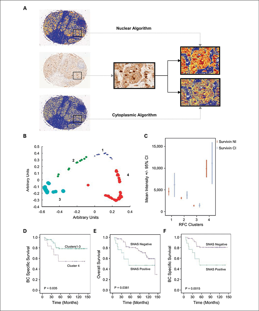
Purpose: Survivin (BIRC5) is a promising tumor biomarker. Conflicting data exist on its prognostic effect in breast cancer. These data may at least be partly due to the manual interpretation of immunohistochemical staining, especially as survivin can be located in both the nucleus and cytoplasm. Quantitative determination of survivin expression using image analysis offers the opportunity to develop alternative scoring models for survivin immunohistochemistry. Here, we present such a model.
Experimental design: A breast cancer tissue microarray containing 102 tumors was stained with an anti-survivin antibody. Whole-slide scanning was used to capture high-resolution images. These images were analyzed using automated algorithms to quantify the staining.
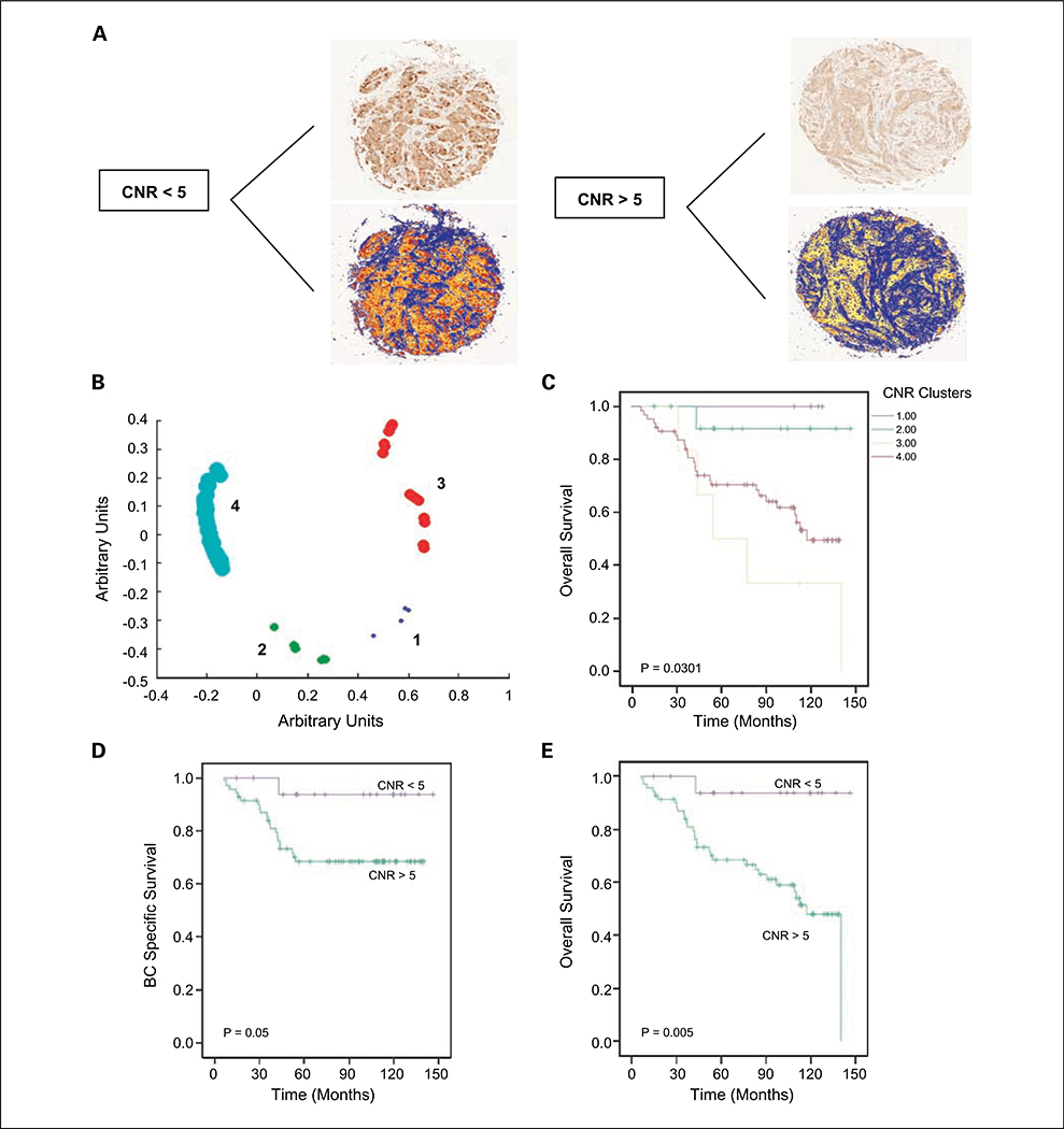
Results: Increased nuclear, but not cytoplasmic, survivin was associated with a reduced overall survival (OS; P = 0.038) and disease-specific survival (P = 0.0015). A high cytoplasmic-to-nuclear ratio (CNR) of survivin was associated with improved OS (P = 0.005) and disease-specific survival (P = 0.05). Multivariate analysis revealed that the survivin CNR was an independent predictor of OS (hazard ratio, 0.09; 95% confidence interval, 0.01-0.76; P = 0.027). A survivin CNR of >5 correlated positively with estrogen receptor (P = 0.019) and progesterone receptor (P = 0.033) levels, whereas it was negatively associated with Ki-67 expression (P = 0.04), p53 status (P = 0.005), and c-myc amplification (P = 0.016).
Conclusion: Different prognostic information is supplied by nuclear and cytoplasmic survivin in breast cancer. Nuclear survivin is a poor prognostic marker in breast cancer. Moreover, CNR of survivin, as determined by image analysis, is an independent prognostic factor.
Figures

References
-
- Duffy MJ, O’Donovan N, Brennan DJ, Gallagher WM, Ryan BM. Survivin: a promising tumor biomarker. Cancer Lett 2007;249:49–60. - PubMed
-
- Velculescu VE, Madden SL, Zhang L, et al. Analysis of human transcriptomes. Nat Genet 1999;23:387–8. - PubMed
-
- Ambrosini G, Adida C, Altieri DC. A novel antiapoptosis gene, survivin, expressed in cancer and lymphoma. Nat Med 1997;3:917–21. - PubMed
-
- Chu JS, Shew JY, Huang CS. Immunohistochemical analysis of survivin expression in primary breast cancers. J Formos Med Assoc 2004;103:925–31. - PubMed
-
- O’Driscoll L, Linehan R, Kennedy SM, et al. Lack of prognostic significance of survivin, survivin-ΔEx3, survivin-2B, galectin-3, bag-1, bax-α and MRP-1 mRNAs in breast cancer. Cancer Lett 2003;201: 225–36. - PubMed
Publication types
MeSH terms
Substances
Grants and funding
LinkOut - more resources
Full Text Sources
Other Literature Sources
Medical
Research Materials
Miscellaneous
