Review
. 2008 Jun;29(4):441-64.
doi: 10.1210/er.2008-0002. Epub 2008 May 1.

Osteoporosis in men

Affiliations
Review

Osteoporosis in men

Sundeep Khosla et al. Endocr Rev. 2008 Jun.

Abstract

With the aging of the population, there is a growing recognition that osteoporosis and fractures in men are a significant public health problem, and both hip and vertebral fractures are associated with increased morbidity and mortality in men. Osteoporosis in men is a heterogeneous clinical entity: whereas most men experience bone loss with aging, some men develop osteoporosis at a relatively young age, often for unexplained reasons (idiopathic osteoporosis). Declining sex steroid levels and other hormonal changes likely contribute to age-related bone loss, as do impairments in osteoblast number and/or activity. Secondary causes of osteoporosis also play a significant role in pathogenesis. Although there is ongoing controversy regarding whether osteoporosis in men should be diagnosed based on female- or male-specific reference ranges (because some evidence indicates that the risk of fracture is similar in women and men for a given level of bone mineral density), a diagnosis of osteoporosis in men is generally made based on male-specific reference ranges. Treatment consists both of nonpharmacological (lifestyle factors, calcium and vitamin D supplementation) and pharmacological (most commonly bisphosphonates or PTH) approaches, with efficacy similar to that seen in women. Increasing awareness of osteoporosis in men among physicians and the lay public is critical for the prevention of fractures in our aging male population.


Figures

Figure 1
Figure 1
Age- and gender-specific incidence of fractures at any site among 5 million adults registered in the General Practice Research Database of the UK, 1988–1998. [Reproduced from T. P. van Staa et al.: Bone 29:517–522, 2001 (15), with permission from Elsevier.]
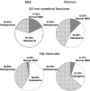
Figure 2
Figure 2
Percentage of nonvertebral or hip fractures that occurred in men and women with osteoporosis, osteopenia, or normal BMD using gender-specific T-scores. [Reproduced from S. C. E. Schuit et al.: Bone 34:195–202, 2004 (20), with permission from Elsevier.]
Figure 3
Figure 3
Patterns of osteopenia and osteoporosis by race or ethnicity in men using either male or female cutoffs compared with the patterns seen in women. NHW, Non-Hispanic white; NHB, non-Hispanic black; MA, Mexican American. [Reproduced from A. C. Looker et al.: J Bone Miner Res 12:1761–1768, 1997 (100), with permission of the American Society for Bone and Mineral Research.]
Figure 4
Figure 4
A, Values for vBMD (mg/cm3) of the total vertebral body in a population sample of Rochester, Minnesota, women and men between the ages of 20 and 97 yr. Individual values and smoother lines are given for premenopausal women in red, for postmenopausal women in blue, and for men in black. B, Values for cortical vBMD at the distal radius in the same cohort. Color code is as in panel A. All changes with age were significant (P < 0.05). [Reproduced from B. L. Riggs et al.: J Bone Miner Res 19:1945–1954, 2004 (109), with permission of the American Society for Bone and Mineral Research.]
Figure 5
Figure 5
A, Values for total area of the femoral neck, adjusted for height, in a population sample between 20 and 97 yr of age. Individual values and smoother lines are given for premenopausal women in red, for postmenopausal women in blue, and for men in black. B, Values for total marrow area, a surrogate for cortical bone loss caused by endocortical resorption, of the femoral neck, adjusted for height in the sample. Color code is as in panel A. [Reproduced from B. L. Riggs et al.: J Bone Miner Res 19:1945–1954, 2004 (109), with permission of the American Society for Bone and Mineral Research.]
Figure 6
Figure 6
Age-specific rates of change in vBMD at trabecular scanning sites in men at the distal radius (A), distal tibia (B), and lumbar spine (C). Data are shown with a smoothing spline and the 95% confidence interval. [Reproduced from B. L. Riggs et al.: J Bone Miner Res 23:205–214, 2008 (110), with permission of the American Society for Bone and Mineral Research.]
Figure 7
Figure 7
Age-specific changes in vBMD at cortical scanning sites at distal radius (A) and distal tibia (B) in men. Data are shown with a smoothing spline and the 95% confidence interval. [Reproduced from B. L. Riggs et al.: J Bone Miner Res 23:205–214, 2008 (110), with permission of the American Society for Bone and Mineral Research.]
Figure 8
Figure 8
Age-related changes in trabecular bone microstructural variables at the wrist in Rochester, Minnesota, women and men. A, BV/TV; B, TbN; C, TbTh; and D, TbSp. Individual values and smoother lines are given for women using open circles and dashed lines and for men using closed circles and solid lines. [Reproduced from S. Khosla et al.: J Bone Miner Res 21:124–131, 2006 (116), with permission of the American Society for Bone and Mineral Research.]
Figure 9
Figure 9
Percent changes in bone resorption markers (urinary Dpd and NTx) (A) and bone formation markers [serum osteocalcin and N-terminal extension peptide of type I collagen (PINP)] (B) in a group of elderly men (mean age 68 yr) made acutely hypogonadal and treated with an aromatase inhibitor (group A), estrogen alone (group B), testosterone alone (group C), or both (group D). See text for details. Asterisks indicate significance for change from baseline: *, P < 0.05; **, P < 0.01; ***, P < 0.001. [Adapted from A. Falahati-Nini et al.: J Clin Invest 106:1553–1560, 2000 (137), with permission from the American Society of Clinical Investigation.]
Figure 10
Figure 10
A general scheme for the evaluation of men suspected of having increased fracture risk.
Figure 11
Figure 11
Three-year risk of fracture (and 95% confidence limits) by sex-specific total hip BMD T-score and age in older women and men. T-scores for males using male normal values for the total hip are equivalent to the following BMD values: T-score of −2 = 0.753 g/cm2; T-score of −1 = 0.897 g/cm2; T-score of 0 = 1.041 g/cm2. T-scores for females using female normal values for the total hip are equivalent to the following BMD values: T-score of −2 = 0.698 g/cm2; T-score of −1 = 0.820 g/cm2; T-score of 0 = 0.942 g/cm2. T-scores for both sexes using female normal values for the total hip are equivalent to the following BMD values: T-score of −2 = 0.698 g/cm2; T-score of −1 = 0.820 g/cm2; T-score of 0 = 0.942 g/cm2. [Reproduced from S. R. Cummings et al.: J Bone Miner Res 21:1550–1556, 2006 (202), with permission of the American Society for Bone and Mineral Research.]

References

    1. Hannan MT, Felson DT, Dawson-Hughes B, Tucker KL, Cupples LA, Wilson PWF, Kiel DP 2000 Risk factors for longitudinal bone loss in elderly men and women: the Framingham osteoporosis study. J Bone Miner Res 15:710–720 - PubMed
    1. Jones G, Nguyen T, Sambrook P, Kelly PJ, Eisman JA 1994 Progressive loss of bone in the femoral neck in elderly people: longitudinal findings from the Dubbo osteoporosis epidemiology study. BMJ 309:691–695 - PMC - PubMed
    1. Melton LJ, Chrischilles EA, Cooper C, Lane AW, Riggs BL 1992 How many women have osteoporosis. J Bone Miner Res 7:1005–1010 - PubMed
    1. Forsen L, Sogaard AJ, Meyer HE, Edna T, Kopjar B 1999 Survival after hip fracture: short- and long-term excess mortality according to age and gender. Osteoporos Int 10:73–78 - PubMed
    1. Center JR, Nguyen TV, Schneider D, Sambrook PN, Eisman JA 1999 Mortality after all major types of osteoporotic fracture in men and women: an observational study. Lancet 353:878–882 - PubMed

Publication types

Substances