Review
. 2008 Jun;29(4):403-40.
doi: 10.1210/er.2007-0038. Epub 2008 May 1.

Osteoimmunology: interactions of the bone and immune system

Affiliations
Review

Osteoimmunology: interactions of the bone and immune system

Joseph Lorenzo et al. Endocr Rev. 2008 Jun.

Abstract

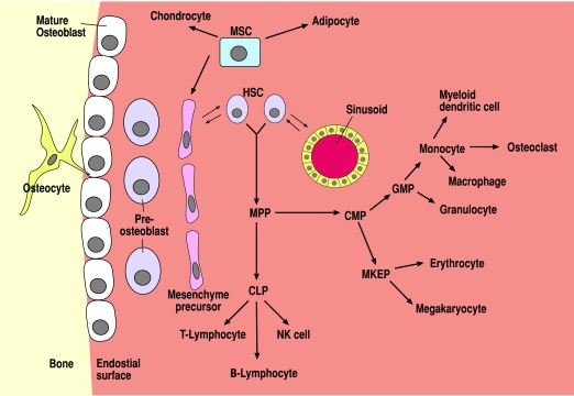
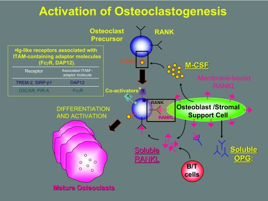
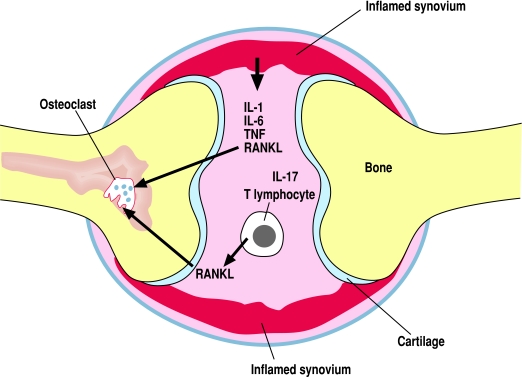
Bone and the immune system are both complex tissues that respectively regulate the skeleton and the body's response to invading pathogens. It has now become clear that these organ systems often interact in their function. This is particularly true for the development of immune cells in the bone marrow and for the function of bone cells in health and disease. Because these two disciplines developed independently, investigators in each don't always fully appreciate the significance that the other system has on the function of the tissue they are studying. This review is meant to provide a broad overview of the many ways that bone and immune cells interact so that a better understanding of the role that each plays in the development and function of the other can develop. It is hoped that an appreciation of the interactions of these two organ systems will lead to better therapeutics for diseases that affect either or both.


Figures

Figure 1
Figure 1
Scheme for the interactions of osteoblasts with hematopoiesis. HSCs reside in the bone marrow adjacent to either osteoblast lineage cells or sinusoids. Both of these likely produce signals that control HSC replication and differentiation. HSC can remain dormant or replicate to either self-renew or differentiate into multipotential progenitor (MPP) cells. MPP can differentiate into either common lymphoid progenitors (CLP), which have the capacity to differentiate into precursors for T-lymphocytes, B-lymphocytes or natural killer (NK) cells; or MPP can become common myeloid precursor (CMP) cells, which are the precursor cells for all other hematopoietic lineages. CMP can differentiate into either granulocyte-macrophage progenitor (GMP) cells or megakaryocyte-erythroid progenitor (MKEP) cells. In turn, MKEP can differentiate into either erythrocytes or megakaryocytes. GMP can differentiate into monocytes or granulocytes. Bone marrow monocytes are precursors for myeloid dendritic cells, macrophages, and osteoclasts. Osteoblasts derive from a mesenchymal precursor cell (MSC) that is multipotential and can also differentiate into chondrocytes and adipocytes. Like the hematopoietic system, differentiation of MSC toward the osteoblast lineage involves multiple intermediates including mesenchyme precursors, preosteoblasts, and mature (matrix producing) osteoblasts. Finally, some mature osteoblasts appear to differentiate further into osteocytes, which are encased in the mineralized matrix of bone. [Derived from Ref. .]
Figure 2
Figure 2
Activation of osteoclastogenesis. Osteoclast precursor cells replicate and are induced to express RANK when stimulated by the binding of M-CSF to its receptor c-fms. In states in which osteoclastogenesis is stimulated, osteoblast or stromal support cells express relatively more RANKL than OPG. This facilitates the binding of RANKL to RANK, which is the critical signal for the differentiation of mature osteoclasts from precursor cells. However, the formation of mature osteoclasts is significantly enhanced by costimulatory molecules on osteoclast precursor cells. It is critical that the ITAM proteins DAP12 and FcγR on the surface of osteoclast precursor cells interact with their respective Ig-like receptors (TREM-2 and SIRP-β1 with DAP12; OSCAR and PIR-A with FcγR) for costimulation to occur. In addition, in inflammatory states and, possibly, in normal physiology, B and T lymphocytes also produce RANKL, which can influence osteoclastogenesis.
Figure 3
Figure 3
Regulation of osteoclastogenesis in inflammation. In inflammatory states such as inflammatory arthritis, local production of proinflammatory cytokines (IL-1, IL-6, and TNF) as well as RANKL by inflamed tissues such as the synovium leads to stimulation of osteoclastogenesis and bone destruction. In addition, IL-17-producing TH17 T lymphocytes stimulate local production of RANKL by inflamed tissues and produce RANKL themselves, which enhances resorptive destruction of bone at sites adjacent to the inflammation.

Similar articles

Cited by

References

    1. Raisz LG, Kream BE, Lorenzo JA, Larsen PR, Kronenberg HM, Melmed S, Polonsky KS 2002 Metabolic bone disease. In: Davies TF, Larsen PR, Kronenberg HM, eds. Williams textbook of endocrinology. Philadelphia: W.B. Saunders; 1373
    1. Rosenberg HF, Gallin JI, Paul WE 1999 Inflammation. In: Paul WE, ed. Fundamental immunology. Philadelphia: Lippincott-Raven; 1051
    1. Alnaeeli M, Penninger JM, Teng YT 2006 Immune interactions with CD4+ T cells promote the development of functional osteoclasts from murine CD11c+ dendritic cells. J Immunol 177:3314–3326 - PubMed
    1. Horowitz MC, Lorenzo J, Bilezikian J, Raisz L, Rodan G 1996 Local regulators of bone: IL-1, TNF, and lymphotoxin, interferon γ, IL-8, IL-10, IL-4, the LIF/IL-6 family and additional cytokines. In: Bilezikian JP, Raisz LG, Rodan GA, eds. Principals of bone biology, 2nd ed. San Diego: Academic Press; 961–977
    1. Raisz LG 1981 What marrow does to bone. N Engl J Med 304:1485–1486 - PubMed

Publication types