

In vivo imaging of membrane-associated glycans in developing zebrafish
- PMID: 18451302
- PMCID: PMC2701225
- DOI: 10.1126/science.1155106
In vivo imaging of membrane-associated glycans in developing zebrafish
Abstract
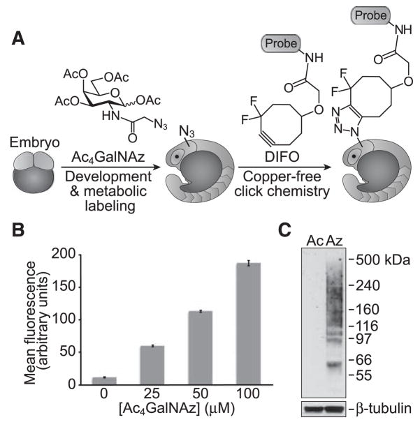
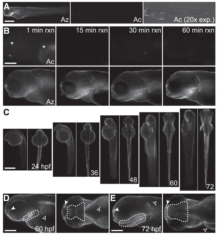
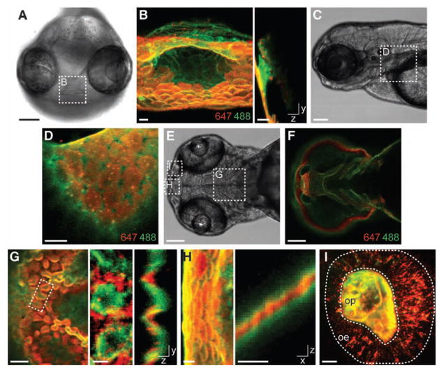
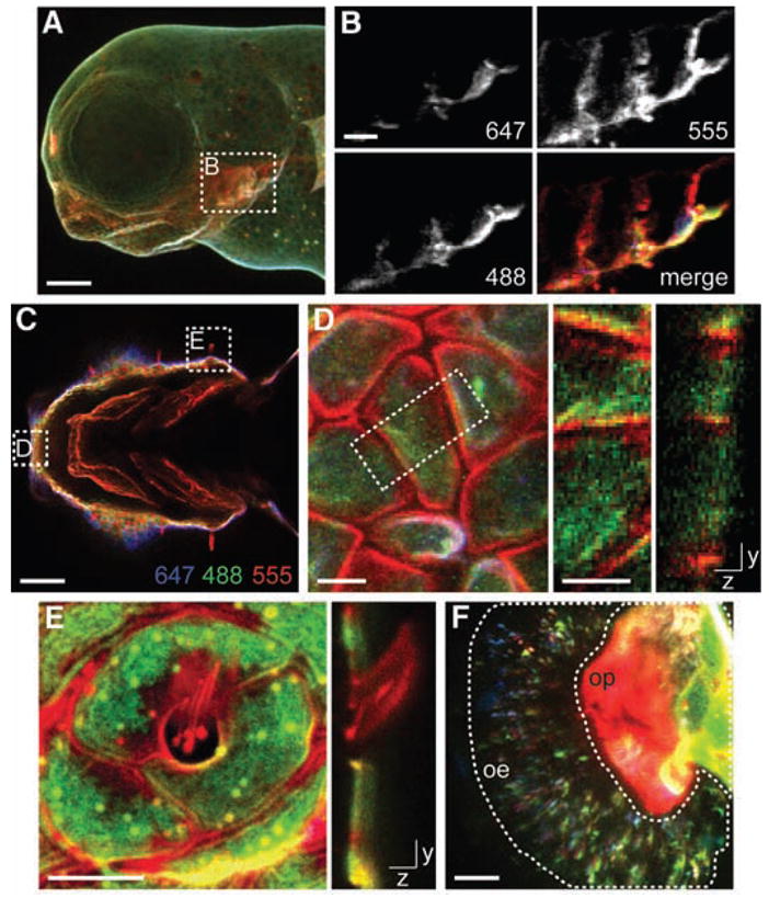
Glycans are attractive targets for molecular imaging but have been inaccessible because of their incompatibility with genetically encoded reporters. We demonstrated the noninvasive imaging of glycans in live developing zebrafish, using a chemical reporter strategy. Zebrafish embryos were treated with an unnatural sugar to metabolically label their cell-surface glycans with azides. Subsequently, the embryos were reacted with fluorophore conjugates by means of copper-free click chemistry, enabling the visualization of glycans in vivo at subcellular resolution during development. At 60 hours after fertilization, we observed an increase in de novo glycan biosynthesis in the jaw region, pectoral fins, and olfactory organs. Using a multicolor detection strategy, we performed a spatiotemporal analysis of glycan expression and trafficking and identified patterns that would be undetectable with conventional molecular imaging approaches.
Figures
References
-
- Haltiwanger RS, Lowe JB. Annu Rev Biochem. 2004;73:491. - PubMed
-
- Ohtsubo K, Marth JD. Cell. 2006;126:855. - PubMed
-
- Hebert DN, Garman SC, Molinari M. Trends Cell Biol. 2005;15:364. - PubMed
-
- Wopereis S, Lefeber DJ, Morava E, Wevers RA. Clin Chem. 2006;52:574. - PubMed
-
- Prescher JA, Bertozzi CR. Nat Chem Biol. 2005;1:13. - PubMed
Publication types
MeSH terms
Substances
Grants and funding
LinkOut - more resources
Full Text Sources
Other Literature Sources
