

Phosphorylation by p38 MAPK as an alternative pathway for GSK3beta inactivation
- PMID: 18451303
- PMCID: PMC2597039
- DOI: 10.1126/science.1156037
Phosphorylation by p38 MAPK as an alternative pathway for GSK3beta inactivation
Abstract
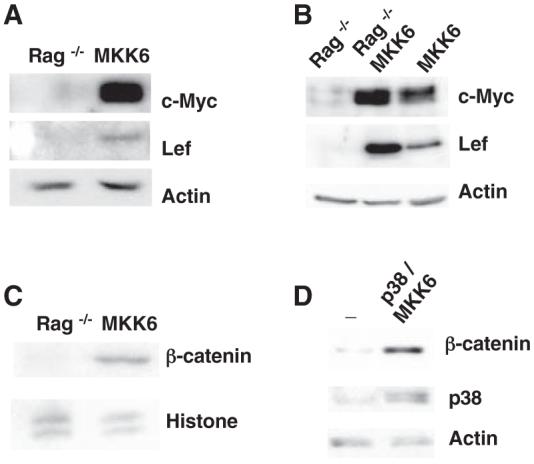
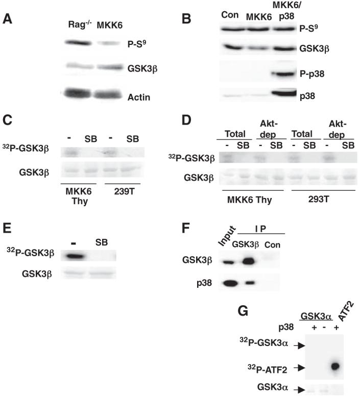
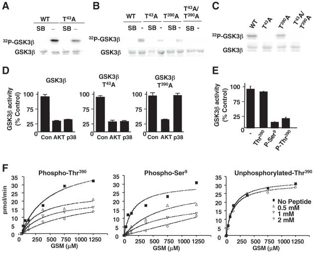
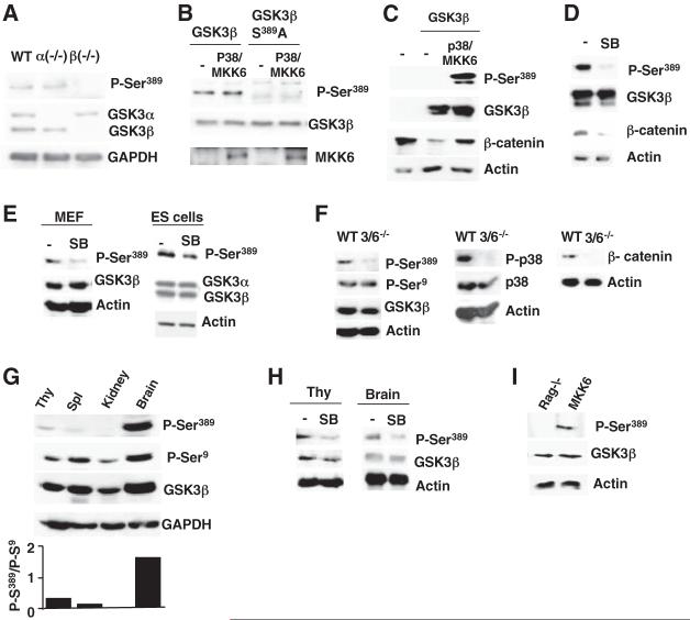
Glycogen synthase kinase 3beta (GSK3beta) is involved in metabolism, neurodegeneration, and cancer. Inhibition of GSK3beta activity is the primary mechanism that regulates this widely expressed active kinase. Although the protein kinase Akt inhibits GSK3beta by phosphorylation at the N terminus, preventing Akt-mediated phosphorylation does not affect the cell-survival pathway activated through the GSK3beta substrate beta-catenin. Here, we show that p38 mitogen-activated protein kinase (MAPK) also inactivates GSK3beta by direct phosphorylation at its C terminus, and this inactivation can lead to an accumulation of beta-catenin. p38 MAPK-mediated phosphorylation of GSK3beta occurs primarily in the brain and thymocytes. Activation of beta-catenin-mediated signaling through GSK3beta inhibition provides a potential mechanism for p38 MAPK-mediated survival in specific tissues.
Figures
Similar articles
-
Upregulation of RGS4 expression by IL-1beta in colonic smooth muscle is enhanced by ERK1/2 and p38 MAPK and inhibited by the PI3K/Akt/GSK3beta pathway.Am J Physiol Cell Physiol. 2009 Jun;296(6):C1310-20. doi: 10.1152/ajpcell.00573.2008. Epub 2009 Apr 15. Am J Physiol Cell Physiol. 2009. PMID: 19369446 Free PMC article.
-
p38 mitogen-activated protein kinase regulates canonical Wnt-beta-catenin signaling by inactivation of GSK3beta.J Cell Sci. 2008 Nov 1;121(Pt 21):3598-607. doi: 10.1242/jcs.032854. J Cell Sci. 2008. PMID: 18946023
-
Epidermal growth factor-stimulated extravillous cytotrophoblast motility is mediated by the activation of PI3-K, Akt and both p38 and p42/44 mitogen-activated protein kinases.Hum Reprod. 2008 Aug;23(8):1733-41. doi: 10.1093/humrep/den178. Epub 2008 May 16. Hum Reprod. 2008. PMID: 18487214 Free PMC article.
-
Mechanical regulation of glycogen synthase kinase 3β (GSK3β) in mesenchymal stem cells is dependent on Akt protein serine 473 phosphorylation via mTORC2 protein.J Biol Chem. 2011 Nov 11;286(45):39450-6. doi: 10.1074/jbc.M111.265330. Epub 2011 Sep 28. J Biol Chem. 2011. PMID: 21956113 Free PMC article.
-
Proteasome inhibition-induced p38 MAPK/ERK signaling regulates autophagy and apoptosis through the dual phosphorylation of glycogen synthase kinase 3β.Biochem Biophys Res Commun. 2012 Feb 24;418(4):759-64. doi: 10.1016/j.bbrc.2012.01.095. Epub 2012 Jan 28. Biochem Biophys Res Commun. 2012. PMID: 22310719
Cited by
-
Glycogen synthase kinase 3: a point of integration in Alzheimer's disease and a therapeutic target?Int J Alzheimers Dis. 2012;2012:276803. doi: 10.1155/2012/276803. Epub 2012 Jun 14. Int J Alzheimers Dis. 2012. PMID: 22779025 Free PMC article.
-
Regulation of onco and tumor suppressor MiRNAs by mTORC1 inhibitor PRP-1 in human chondrosarcoma.Tumour Biol. 2014 Mar;35(3):2335-41. doi: 10.1007/s13277-013-1309-7. Epub 2013 Nov 1. Tumour Biol. 2014. PMID: 24178909
-
Targeting Src-mediated Tyr216 phosphorylation and activation of GSK-3 in prostate cancer cells inhibit prostate cancer progression in vitro and in vivo.Oncotarget. 2014 Feb 15;5(3):775-87. doi: 10.18632/oncotarget.1770. Oncotarget. 2014. PMID: 24519956 Free PMC article.
-
Non-classical p38 map kinase functions: cell cycle checkpoints and survival.Int J Biol Sci. 2009;5(1):44-51. doi: 10.7150/ijbs.5.44. Epub 2008 Dec 19. Int J Biol Sci. 2009. PMID: 19159010 Free PMC article. Review.
-
Nrf2 and Heme Oxygenase-1 Involvement in Atherosclerosis Related Oxidative Stress.Antioxidants (Basel). 2021 Sep 14;10(9):1463. doi: 10.3390/antiox10091463. Antioxidants (Basel). 2021. PMID: 34573095 Free PMC article. Review.
References
Publication types
MeSH terms
Substances
Grants and funding
LinkOut - more resources
Full Text Sources
Other Literature Sources
Molecular Biology Databases
Miscellaneous
