

Mertk receptor mutation reduces efferocytosis efficiency and promotes apoptotic cell accumulation and plaque necrosis in atherosclerotic lesions of apoe-/- mice
- PMID: 18451332
- PMCID: PMC2575060
- DOI: 10.1161/ATVBAHA.108.167197
Mertk receptor mutation reduces efferocytosis efficiency and promotes apoptotic cell accumulation and plaque necrosis in atherosclerotic lesions of apoe-/- mice
Abstract
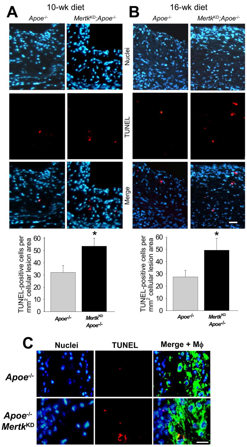
Objective: Atherosclerotic plaques that are prone to disruption and acute thrombotic vascular events are characterized by large necrotic cores. Necrotic cores result from the combination of macrophage apoptosis and defective phagocytic clearance (efferocytosis) of these apoptotic cells. We previously showed that macrophages with tyrosine kinase-defective Mertk receptor (Mertk(KD)) have a defect in phagocytic clearance of apoptotic macrophages in vitro. Herein we test the hypothesis that the Mertk(KD) mutation would result in increased accumulation of apoptotic cells and promote necrotic core expansion in a mouse model of advanced atherosclerosis.
Methods and results: Mertk(KD);Apoe(-/-) mice and control Apoe(-/-) mice were fed a Western-type diet for 10 or 16 weeks, and aortic root lesions were analyzed for apoptosis and plaque necrosis. We found that the plaques of the Mertk(KD);Apoe(-/-) mice had a significant increase in terminal deoxynucleotidyl transferase-mediated dUTP nick end-labeling (TUNEL)-positive apoptotic cells. Most importantly, there were more non-macrophage-associated apoptotic cells in the Mertk(KD) lesions, consistent with defective efferocytosis. The more advanced (16-week) Mertk(KD);Apoe(-/-) plaques were more necrotic, consistent with a progression from apoptotic cell accumulation to plaque necrosis in the setting of a defective efferocytosis receptor.
Conclusions: In a mouse model of advanced atherosclerosis, mutation of the phagocytic Mertk receptor promotes the accumulation of apoptotic cells and the formation of necrotic plaques. These data are consistent with the notion that a defect in an efferocytosis receptor can accelerate the progression of atherosclerosis and suggest a novel therapeutic target to prevent advanced plaque progression and its clinical consequences.
Figures


Comment in
-
Macrophage function and its impact on atherosclerotic lesion composition, progression, and stability: the good, the bad, and the ugly.Arterioscler Thromb Vasc Biol. 2008 Aug;28(8):1413-5. doi: 10.1161/ATVBAHA.108.169144. Arterioscler Thromb Vasc Biol. 2008. PMID: 18650503 No abstract available.
References
-
- Virmani R, Burke AP, Kolodgie FD, Farb A. Vulnerable plaque: the pathology of unstable coronary lesions. J Interv Cardiol. 2002;15:439–446. - PubMed
-
- Tabas I. Consequences and therapeutic implications of macrophage apoptosis in atherosclerosis: the importance of lesion stage and phagocytic efficiency. Arterioscler Thromb Vasc Biol. 2005;25:2255–2264. - PubMed
-
- Schrijvers DM, De Meyer GR, Herman AG, Martinet W. Phagocytosis in atherosclerosis: Molecular mechanisms and implications for plaque progression and stability. Cardiovasc Res. 2007;73:470–480. - PubMed
-
- Vandivier RW, Henson PM, Douglas IS. Burying the dead: the impact of failed apoptotic cell removal (efferocytosis) on chronic inflammatory lung disease. Chest. 2006;129:1673–1682. - PubMed
-
- Tabas I. Mouse models of apoptosis and efferocytosis. Curr Drug Targets. 2008 - PubMed
Publication types
MeSH terms
Substances
Grants and funding
LinkOut - more resources
Full Text Sources
Medical
Molecular Biology Databases
Miscellaneous
