

Myocardin is sufficient for a smooth muscle-like contractile phenotype
- PMID: 18451334
- PMCID: PMC2574857
- DOI: 10.1161/ATVBAHA.108.166066
Myocardin is sufficient for a smooth muscle-like contractile phenotype
Abstract
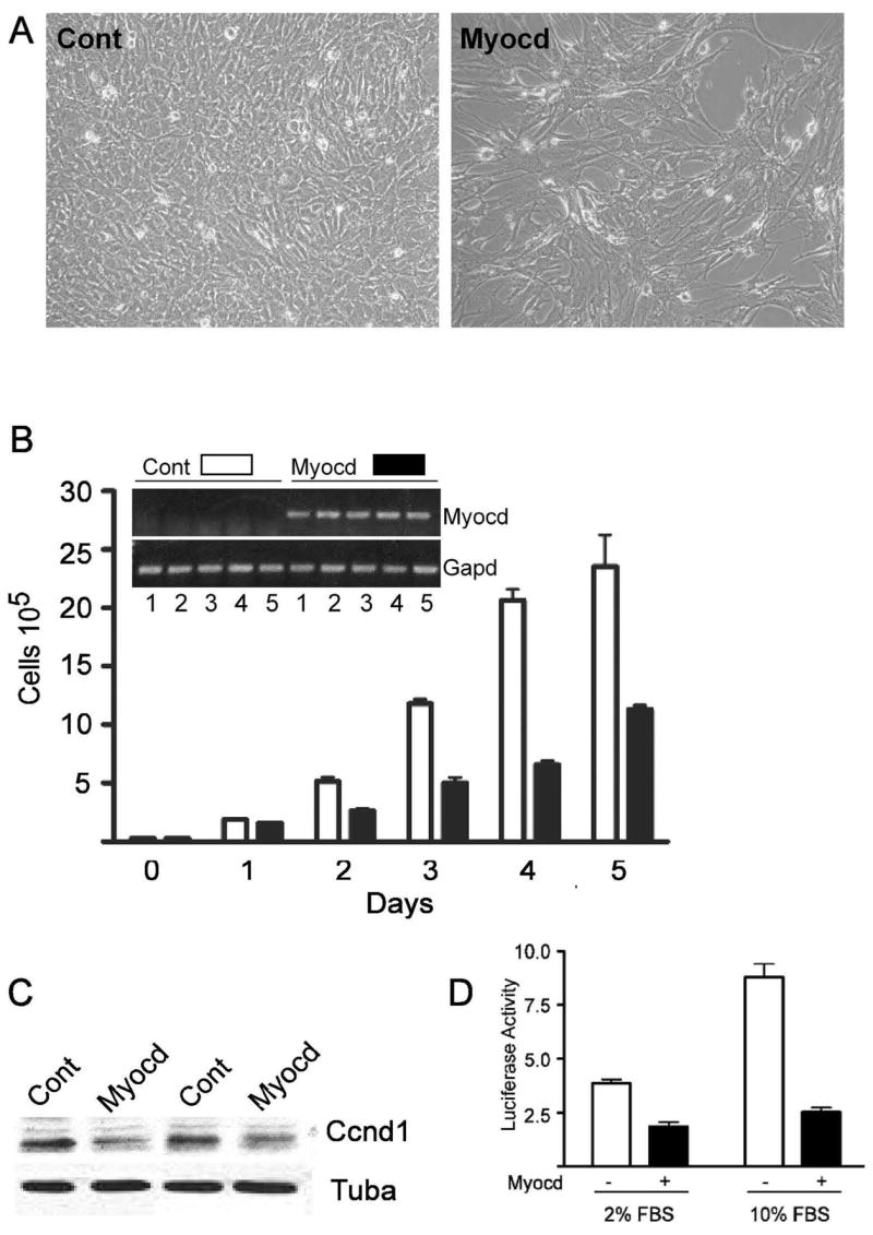
Background: Myocardin (Myocd) is a strong coactivator that binds the serum response factor (SRF) transcription factor over CArG elements embedded within smooth muscle cell (SMC) and cardiac muscle cyto-contractile genes. Here, we sought to ascertain whether Myocd-mediated gene expression confers a structural and physiological cardiac or SMC phenotype.
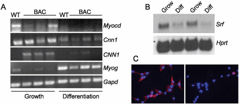
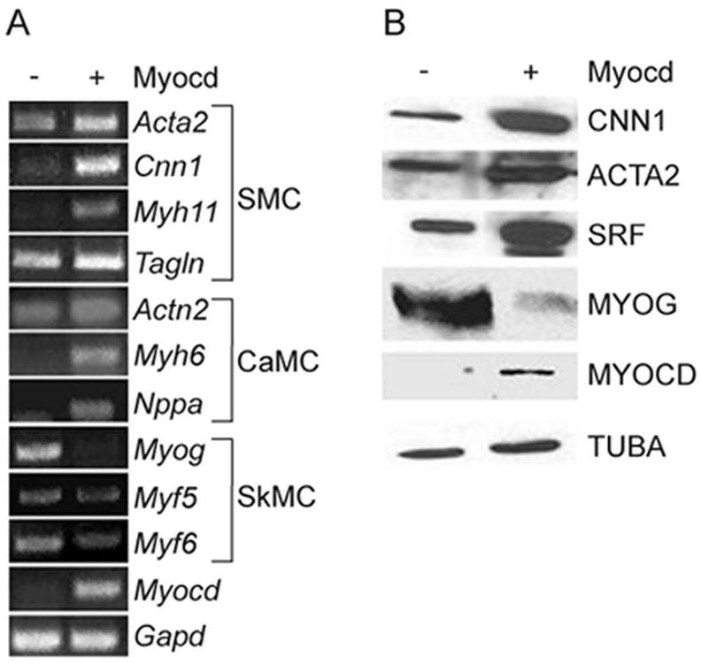
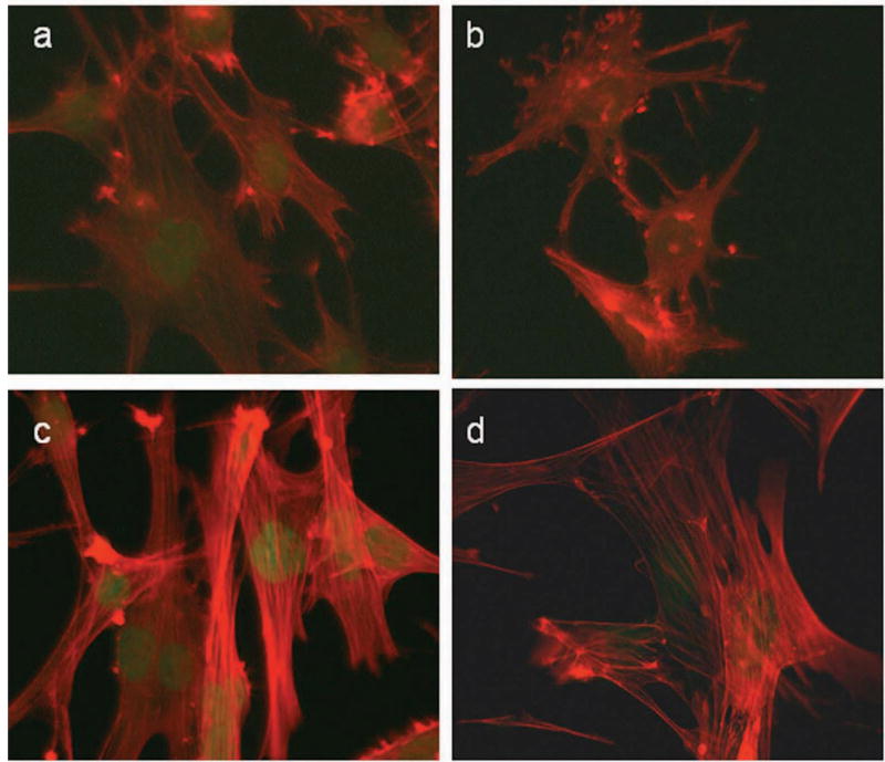
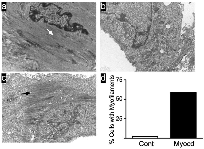
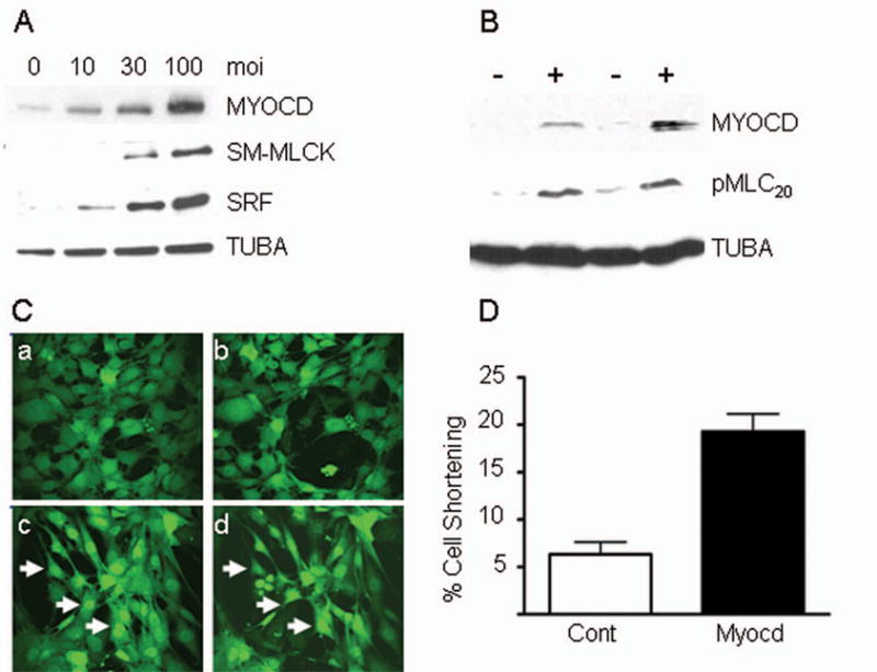
Methods and results: Adenoviral-mediated expression of Myocd in the BC(3)H1 cell line induces cardiac and SMC genes while suppressing both skeletal muscle markers and cell growth. Immunofluorescence microscopy shows that SRF and a SMC-like cyto-contractile apparatus are elevated with Myocd overexpression. A short hairpin RNA to Srf impairs BC(3)H1 cyto-architecture; however, cotransduction with Myocd results in complete restoration of the cyto-architecture. Electron microscopic studies demonstrate a SMC ultrastructural phenotype with no evidence for cardiac sarcomerogenesis. Biochemical and time-lapsed videomicroscopy assays reveal clear evidence for Myocd-induced SMC-like contraction.
Conclusions: Myocd is sufficient for the establishment of a SMC-like contractile phenotype.
Figures
Comment in
-
Myocardin: dominant driver of the smooth muscle cell contractile phenotype.Arterioscler Thromb Vasc Biol. 2008 Aug;28(8):1416-7. doi: 10.1161/ATVBAHA.108.168930. Arterioscler Thromb Vasc Biol. 2008. PMID: 18650504 Free PMC article. No abstract available.
Similar articles
-
Ten-eleven translocation-2 (TET2) is a master regulator of smooth muscle cell plasticity.Circulation. 2013 Oct 29;128(18):2047-57. doi: 10.1161/CIRCULATIONAHA.113.002887. Epub 2013 Sep 27. Circulation. 2013. PMID: 24077167 Free PMC article.
-
Myocardin-dependent activation of the CArG box-rich smooth muscle gamma-actin gene: preferential utilization of a single CArG element through functional association with the NKX3.1 homeodomain protein.J Biol Chem. 2009 Nov 20;284(47):32582-90. doi: 10.1074/jbc.M109.033910. Epub 2009 Sep 21. J Biol Chem. 2009. PMID: 19797053 Free PMC article.
-
Myocardin regulates expression of contractile genes in smooth muscle cells and is required for closure of the ductus arteriosus in mice.J Clin Invest. 2008 Feb;118(2):515-25. doi: 10.1172/JCI33304. J Clin Invest. 2008. PMID: 18188448 Free PMC article.
-
Myocardin: A novel player in atherosclerosis.Atherosclerosis. 2017 Feb;257:266-278. doi: 10.1016/j.atherosclerosis.2016.12.002. Epub 2016 Dec 1. Atherosclerosis. 2017. PMID: 28012646 Review.
-
Serum response factor: toggling between disparate programs of gene expression.J Mol Cell Cardiol. 2003 Jun;35(6):577-93. doi: 10.1016/s0022-2828(03)00110-x. J Mol Cell Cardiol. 2003. PMID: 12788374 Review.
Cited by
-
Circulating miRNAs: reflecting or affecting cardiovascular disease?Curr Hypertens Rep. 2012 Dec;14(6):498-509. doi: 10.1007/s11906-012-0310-7. Curr Hypertens Rep. 2012. PMID: 22996205 Review.
-
Inducible Prmt1 ablation in adult vascular smooth muscle leads to contractile dysfunction and aortic dissection.Exp Mol Med. 2021 Oct;53(10):1569-1579. doi: 10.1038/s12276-021-00684-x. Epub 2021 Oct 11. Exp Mol Med. 2021. PMID: 34635781 Free PMC article.
-
Transforming growth factor-beta1 (TGF-beta1) utilizes distinct pathways for the transcriptional activation of microRNA 143/145 in human coronary artery smooth muscle cells.J Biol Chem. 2011 Aug 26;286(34):30119-29. doi: 10.1074/jbc.M111.258814. Epub 2011 Jun 28. J Biol Chem. 2011. PMID: 21712382 Free PMC article.
-
Smooth muscle calponin: an unconventional CArG-dependent gene that antagonizes neointimal formation.Arterioscler Thromb Vasc Biol. 2011 Oct;31(10):2172-80. doi: 10.1161/ATVBAHA.111.232785. Epub 2011 Aug 4. Arterioscler Thromb Vasc Biol. 2011. PMID: 21817093 Free PMC article.
-
Vascular smooth muscle cell differentiation-2010.J Biomed Res. 2010 May;24(3):169-80. doi: 10.1016/S1674-8301(10)60026-7. J Biomed Res. 2010. PMID: 23554628 Free PMC article.
References
-
- Spaet TH, Lejnieks I. Mitotic activity of rabbit blood vessels. Proc Soc Exp Biol Med. 1967;125:1197–1201. - PubMed
-
- Owens GK, Kumar MS, Wamhoff BR. Molecular regulation of vascular smooth muscle cell differentiation in development and disease. Physiol Rev. 2004;84:767–801. - PubMed
-
- Ramaswamy S, Ross KN, Lander ES, Golub TR. A molecular signature of metastasis in primary solid tumors. Nat Genet. 2003;33:49–54. - PubMed
Publication types
MeSH terms
Substances
Grants and funding
LinkOut - more resources
Full Text Sources
Molecular Biology Databases
Miscellaneous
