

Isolation and short-term culture of primary keratinocytes, hair follicle populations and dermal cells from newborn mice and keratinocytes from adult mice for in vitro analysis and for grafting to immunodeficient mice
- PMID: 18451788
- PMCID: PMC6299324
- DOI: 10.1038/nprot.2008.50
Isolation and short-term culture of primary keratinocytes, hair follicle populations and dermal cells from newborn mice and keratinocytes from adult mice for in vitro analysis and for grafting to immunodeficient mice
Abstract
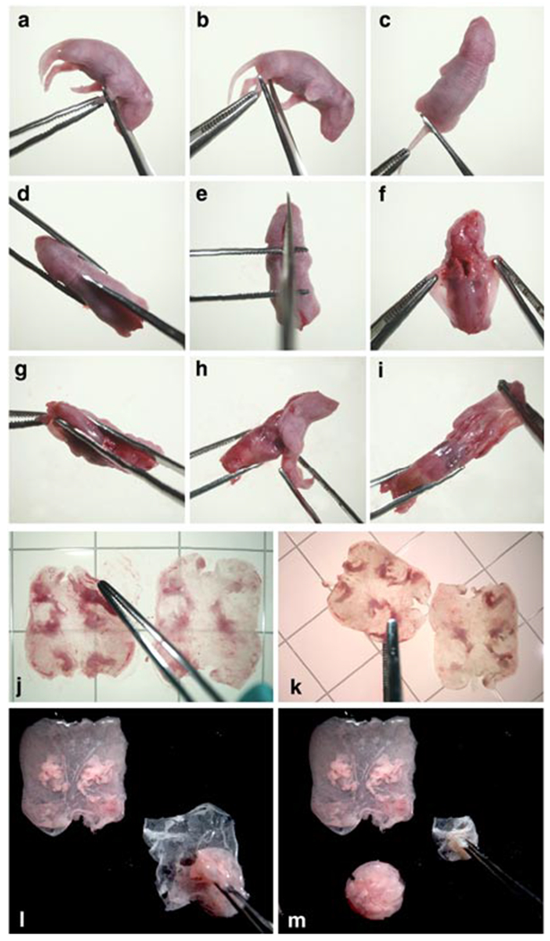
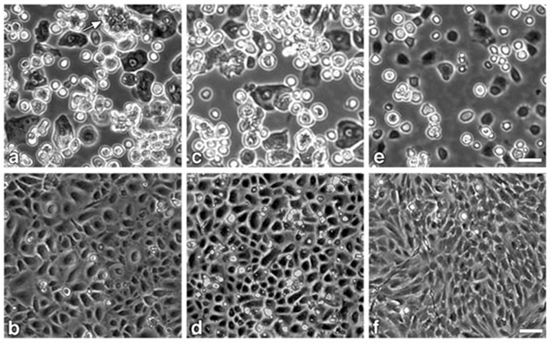
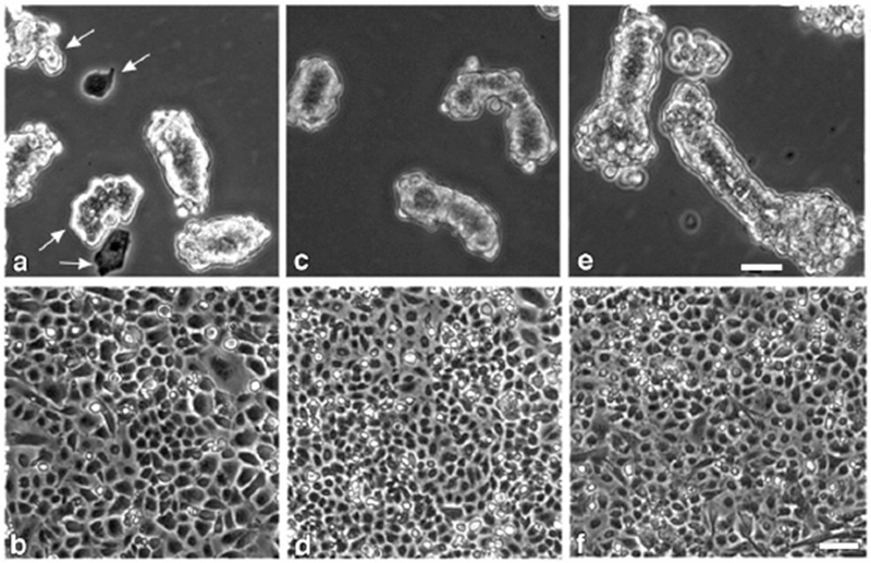
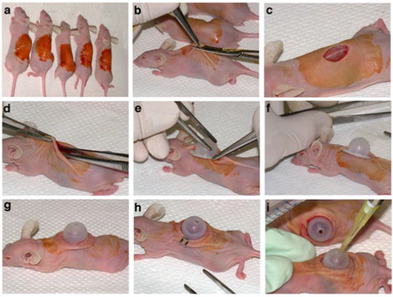
Protocols for preparing and culturing primary keratinocytes from newborn and adult mouse epidermis have evolved over the past 35 years. This protocol is now routinely applied to mice of various genetic backgrounds for in vitro studies of signaling pathways in differentiation and cell transformation, and for assessing the in vivo phenotype of altered keratinocytes in grafts of cells on immunodeficient mice. Crucial in the development and application of the procedure was the observation that keratinocytes proliferate in media of low calcium concentration, but rapidly commit to differentiation at calcium concentrations >0.07 mM after the initial attachment period. Preparing primary keratinocytes from ten newborn mice requires 2-3 h of hands-on time. Related procedures are also provided: preparing immature hair follicle buds, developing dermal hair follicles and fibroblasts from newborn mice, preparing primary keratinocytes from adult mice and grafting cell mixtures on athymic nude mice.
Figures
Similar articles
-
A model system to analyse the ability of human keratinocytes to form hair follicles.Exp Dermatol. 2014 Jun;23(6):443-6. doi: 10.1111/exd.12424. Exp Dermatol. 2014. PMID: 24758480
-
Isolation of a mesenchymal cell population from murine dermis that contains progenitors of multiple cell lineages.FASEB J. 2007 Jul;21(9):2050-63. doi: 10.1096/fj.06-5880com. Epub 2007 Mar 23. FASEB J. 2007. PMID: 17384147 Free PMC article.
-
In vitro expansion of immature melanoblasts and their ability to repopulate melanocyte stem cells in the hair follicle.J Invest Dermatol. 2008 Feb;128(2):408-20. doi: 10.1038/sj.jid.5700997. Epub 2007 Jul 26. J Invest Dermatol. 2008. PMID: 17657242
-
Methods for the isolation and 3D culture of dermal papilla cells from human hair follicles.Exp Dermatol. 2017 Jun;26(6):491-496. doi: 10.1111/exd.13368. Exp Dermatol. 2017. PMID: 28418608 Free PMC article. Review.
-
Strategies to enhance epithelial-mesenchymal interactions for human hair follicle bioengineering.J Dermatol Sci. 2013 May;70(2):78-87. doi: 10.1016/j.jdermsci.2013.02.004. Epub 2013 Feb 28. J Dermatol Sci. 2013. PMID: 23557720 Review.
Cited by
-
PDGFRα signaling drives adipose tissue fibrosis by targeting progenitor cell plasticity.Genes Dev. 2015 Jun 1;29(11):1106-19. doi: 10.1101/gad.260554.115. Epub 2015 May 27. Genes Dev. 2015. PMID: 26019175 Free PMC article.
-
Cancer susceptibility and embryonic lethality in Mob1a/1b double-mutant mice.J Clin Invest. 2012 Dec;122(12):4505-18. doi: 10.1172/JCI63735. Epub 2012 Nov 12. J Clin Invest. 2012. PMID: 23143302 Free PMC article.
-
Filaggrin loss-of-function mutations are associated with enhanced expression of IL-1 cytokines in the stratum corneum of patients with atopic dermatitis and in a murine model of filaggrin deficiency.J Allergy Clin Immunol. 2012 Apr;129(4):1031-9.e1. doi: 10.1016/j.jaci.2011.12.989. Epub 2012 Feb 8. J Allergy Clin Immunol. 2012. PMID: 22322004 Free PMC article.
-
IL-6 Negatively Regulates IL-22Rα Expression on Epidermal Keratinocytes: Implications for Irritant Contact Dermatitis.J Immunol Res. 2019 Oct 29;2019:6276254. doi: 10.1155/2019/6276254. eCollection 2019. J Immunol Res. 2019. PMID: 31781680 Free PMC article.
-
Antimicrobial peptide nisin induces spherical distribution of macropinocytosis-like cytokeratin 5 and cytokeratin 17 following immediate derangement of the cell membrane.Anat Cell Biol. 2022 Jun 30;55(2):190-204. doi: 10.5115/acb.21.168. Epub 2021 Dec 14. Anat Cell Biol. 2022. PMID: 34903675 Free PMC article.
References
-
- Hennings H et al. Calcium regulation of growth and differentiation of mouse epidermal cells in culture. Cell 19, 245–254 (1980). - PubMed
-
- Hennings H Primary culture of keratinocytes from newborn mouse epidermis in medium with lowered levels of Ca2+ In Keratinocyte Methods. (eds.Leigh I & Watt FM) pp. 21–23. (Cambridge University Press, Cambridge, UK, 1994).
-
- Dlugosz AA, Glick AB, Tennenbaum T, Weinberg WC, & Yuspa SH Isolation and utilization of epidermal keratinocytes for oncogene research In Methods in Enzymology, Vol 254, pp. 3–20, (Academic Press, New York, New York, USA, 1995). - PubMed
-
- Kulesz-Martin MF, Koehler B, Hennings H, & Yuspa SH Quantitative assay for carcinogen altered differentiation in mouse epidermal cells. Carcinogenesis 1, 995–1006 (1980). - PubMed
MeSH terms
Substances
Grants and funding
LinkOut - more resources
Full Text Sources
Other Literature Sources
Miscellaneous
