

L-Measure: a web-accessible tool for the analysis, comparison and search of digital reconstructions of neuronal morphologies
- PMID: 18451794
- PMCID: PMC4340709
- DOI: 10.1038/nprot.2008.51
L-Measure: a web-accessible tool for the analysis, comparison and search of digital reconstructions of neuronal morphologies
Abstract
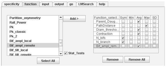
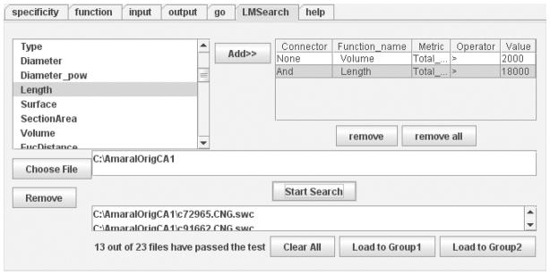
L-Measure (LM) is a freely available software tool for the quantitative characterization of neuronal morphology. LM computes a large number of neuroanatomical parameters from 3D digital reconstruction files starting from and combining a set of core metrics. After more than six years of development and use in the neuroscience community, LM enables the execution of commonly adopted analyses as well as of more advanced functions. This report illustrates several LM protocols: (i) extraction of basic morphological parameters, (ii) computation of frequency distributions, (iii) measurements from user-specified subregions of the neuronal arbors, (iv) statistical comparison between two groups of cells and (v) filtered selections and searches from collections of neurons based on any Boolean combination of the available morphometric measures. These functionalities are easily accessed and deployed through a user-friendly graphical interface and typically execute within few minutes on a set of approximately 20 neurons. The tool is available at http://krasnow.gmu.edu/cn3 for either online use on any Java-enabled browser and platform or download for local execution under Windows and Linux.
Figures
Similar articles
-
A cross-platform freeware tool for digital reconstruction of neuronal arborizations from image stacks.Neuroinformatics. 2005;3(4):343-60. doi: 10.1385/NI:3:4:343. Neuroinformatics. 2005. PMID: 16284416
-
The CARMEN software as a service infrastructure.Philos Trans A Math Phys Eng Sci. 2012 Dec 10;371(1983):20120080. doi: 10.1098/rsta.2012.0080. Print 2013 Jan 28. Philos Trans A Math Phys Eng Sci. 2012. PMID: 23230159
-
Large scale similarity search across digital reconstructions of neural morphology.Neurosci Res. 2022 Aug;181:39-45. doi: 10.1016/j.neures.2022.05.004. Epub 2022 May 14. Neurosci Res. 2022. PMID: 35580795 Free PMC article.
-
Generation, description and storage of dendritic morphology data.Philos Trans R Soc Lond B Biol Sci. 2001 Aug 29;356(1412):1131-45. doi: 10.1098/rstb.2001.0905. Philos Trans R Soc Lond B Biol Sci. 2001. PMID: 11545695 Free PMC article. Review.
-
Successes and rewards in sharing digital reconstructions of neuronal morphology.Neuroinformatics. 2007 Fall;5(3):154-60. doi: 10.1007/s12021-007-0010-7. Neuroinformatics. 2007. PMID: 17917126 Review.
Cited by
-
DSM: Deep sequential model for complete neuronal morphology representation and feature extraction.Patterns (N Y). 2023 Dec 13;5(1):100896. doi: 10.1016/j.patter.2023.100896. eCollection 2024 Jan 12. Patterns (N Y). 2023. PMID: 38264721 Free PMC article.
-
Multiscale Analysis of Neurite Orientation and Spatial Organization in Neuronal Images.Neuroinformatics. 2016 Oct;14(4):465-77. doi: 10.1007/s12021-016-9306-9. Neuroinformatics. 2016. PMID: 27369547 Free PMC article.
-
Machine learning classification reveals robust morphometric biomarker of glial and neuronal arbors.J Neurosci Res. 2023 Jan;101(1):112-129. doi: 10.1002/jnr.25131. Epub 2022 Oct 5. J Neurosci Res. 2023. PMID: 36196621 Free PMC article.
-
Functional Neuroanatomy of the Rat Nucleus Incertus-Medial Septum Tract: Implications for the Cell-Specific Control of the Septohippocampal Pathway.Front Cell Neurosci. 2022 Feb 25;16:836116. doi: 10.3389/fncel.2022.836116. eCollection 2022. Front Cell Neurosci. 2022. PMID: 35281300 Free PMC article.
-
Immunohistochemistry as a detection tool for ion channels involved in dental pain signaling.Saudi Dent J. 2022 Mar;34(3):155-166. doi: 10.1016/j.sdentj.2022.02.004. Epub 2022 Feb 26. Saudi Dent J. 2022. PMID: 35935722 Free PMC article. Review.
References
-
- Pyapali GK, Turner DA. Increased dendritic extent in hippocampal CA1 neurons from aged F344 rats. Neurobiol Aging. 1996;17:601–611. - PubMed
-
- Bulinski JC, et al. Changes in dendritic structure and function following hippocampal lesions: correlations with developmental events? Prog Neurobiol. 1998;55:641–650. - PubMed
-
- Cannon RC, Wheal HV, Turner DA. Dendrites of classes of hippocampal neurons differ in structural complexity and branching patterns. J Comp Neurol. 1999;413:619–633. - PubMed
-
- Ascoli GA. Neuroanatomical algorithms for dendritic modelling. Network. 2002;13:247–260. - PubMed
Publication types
MeSH terms
Grants and funding
LinkOut - more resources
Full Text Sources
