. 2008 Jun 4;27(11):1633-45.
doi: 10.1038/emboj.2008.84. Epub 2008 May 1.

A Wingless and Notch double-repression mechanism regulates G1-S transition in the Drosophila wing

Affiliations

A Wingless and Notch double-repression mechanism regulates G1-S transition in the Drosophila wing

Héctor Herranz et al. EMBO J. .

Abstract

The control of tissue growth and patterning is orchestrated in various multicellular tissues by the coordinated activity of the signalling molecules Wnt/Wingless (Wg) and Notch, and mutations in these pathways can cause cancer. The role of these molecules in the control of cell proliferation and the crosstalk between their corresponding pathways remain poorly understood. Crosstalk between Notch and Wg has been proposed to organize pattern and growth in the Drosophila wing primordium. Here we report that Wg and Notch act in a surprisingly linear pathway to control G1-S progression. We present evidence that these molecules exert their function by regulating the expression of the dmyc proto-oncogene and the bantam micro-RNA, which positively modulated the activity of the E2F transcription factor. Our results demonstrate that Notch acts in this cellular context as a repressor of cell-cycle progression and Wg has a permissive role in alleviating Notch-mediated repression of G1-S progression in wing cells.


Figures
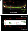
Figure 1
Figure 1
Wg- and Notch-mediated cell interactions involved in DV boundary formation. (A) Late third instar wing imaginal disc labelled to visualize Wg (green) and Senseless (red) protein expression in boundary and non-boundary cells, respectively. (B) Illustration describing the cell interactions that take place between boundary (white) and non-boundary (black) cells that lead to activation of Notch (N) and expression of Wg and Cut along the DV boundary of the Drosophila wing primordium. N, Notch; Dl, Delta; Ser, Serrate.
Figure 2
Figure 2
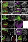
The ZNC is defined by the activity of Notch. Late third instar wing discs labelled to visualize cells in S-phase by BrdU incorporation (antibody to BrdU in red or white (A, C, E, G, I, N, O, P)) or expressing the E2F1-responsive reporter ORC1–GFP (antibody to GFP in pink or white (B, D, F, H, J, Q)). (A, B) Wild-type wing discs labelled to visualize crumbslacZ expression (antibody to β-gal, green). Note failure to incorporate BrdU (A) or low levels of E2F1 activity (B) in those cells expressing high levels of the Notch-regulated gene crumbs, which corresponds to the ZNC. The DV compartment boundary is marked in this and other panels (d, dorsal; v, ventral). (C, D) Notchts2 wing discs reared at the restrictive temperature and labelled to visualize Wg protein expression (green). (E, F) ptcGal4; UASDeltaDN (E) and salGal4; UASmamDN (F) wing discs labelled to visualize Senseless (Sens, green in E) or Wg (green in F) protein expression. Sens expression was used to label the DV boundary. Red lines label the domain of DeltaDN or mamDN expression. (G, H) Wing discs with clones of cells lacking Su(H) activity (Su(H)8), marked by the absence of GFP (G) or β-gal (H) in green. The ZNC is marked by horizontal lines in G. (I) Wing disc with clones of cells lacking arrow activity (arrow2) marked by the absence of GFP. The ZNC is marked by horizontal lines. (J) dppGal4 Gal80ts; UASTCFDN wing disc reared at 29°C during 30 h and labelled to visualize Wg (green) protein expression. The dpp–Gal4 domain is marked by arrowheads. (KR) Wild-type (K, N), C96Gal4; UASTCFDN (L, O), C96Gal4; UAS TCFDN UASNintra (M, P, Q) and C96Gal4; UASGFP (R) wing discs labelled to visualize Cut (green, K–M) or GFP (green, R) protein expression, BrdU incorporation (red, N–P) or activity of the E2F1 responsive reporter ORC1GFP (antibody to GFP in pink, Q). The ZNC is marked by horizontal dashed lines.
Figure 3
Figure 3
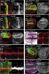
Regulation of E2F activity by Notch and Wg. (AH) AbruptexM1 (A, B), dppGal4;UASNintra (C, C′, D, D′), apGal4 Gal80ts, UASGFP; UASTCFDN wing (E, F), apGal4 Gal80ts, UASGFP; UASAxin (G) and dppGal4 Gal80ts; UASTCFDN (H) late third instar wing discs labelled to visualize cells in S-phase by BrdU incorporation (antibody to BrdU in red or white (A, C, C′, E, G)) or expressing the E2F1-responsive reporter OCT1GFP (antibody to GFP in pink or white (B, D, D′, F, H)). crblacZ (antibody to β-Gal in green (A, B)), Wingless (Wg, green (C, C′, D, D′, H)) and GFP (green (E, G)) protein expression is shown. Panels C′ and D′ show higher magnifications of the discs in panels C and D. The anterior–posterior (ap) boundary is labelled to show the cell-autonomous block in G1 imposed by the activity of N. Nintra activity is labelled by the expression of Wg (red line) and the G1 block by reduced levels of BrdU and dE2F activity. (I, J, K) apGal4, Gal80ts, UASGFP; UASTCFDN (I), wild-type (J) and apGal4 Gal80ts, UASGFP; UASNintra (K) early third instar wing discs labelled to visualize BrdU incorporation (red or white) and GFP (green) expression. The DV compartment boundary is marked in all panels (d, dorsal; v, ventral). Wing discs shown in panels E–I were reared at 29°C during 24 h.
Figure 4
Figure 4
A Wg and Notch double-repression mechanism regulates G1 progression. (AD) dpp–Gal4, UAS–GFP; UASNintraUASE2F (A), ap–Gal4, Gal80ts, UAS–GFP; UAS–TCFDN, UAS–E2F (B), ptc–Gal4, Gal80ts, UAS–GFP; UAS–Nintra, UAS–CycE (C) and ap–Gal4, Gal80ts, UAS–GFP; UAS–TCFDN, UAS–CycE (D) late third instar wing discs labelled to visualize cells in S-phase by BrdU incorporation (antibody to BrdU in red or white) and GFP (green) protein expression. (EG) ap–Gal4, Gal80ts, UAS–GFP; UAS–TCFDN (E) and dpp–Gal4, Gal80ts, UAS–GFP; UAS–TCFDN (F, F′, G) late third instar wing discs labelled to visualize Wingless protein (Wg, red or white, E, G), wg mRNA (F), wglacZ expression (antibody to β-gal, F′) and GFP (green, E) protein expression. Arrowheads in panels F, F′ and G indicate the dpp–gal4 expression stripe. (H) ap–Gal4, Gal80ts, UAS–GFP; UAS–TCFDN, UAS–mamDN late third instar wing disc labelled to visualize cells in S-phase by BrdU incorporation (antibody to BrdU in red or white) and GFP (green) protein expression. (I) Wing disc with clones of cells lacking arrow activity (arrow2), marked by the absence of GFP in green, and labelled to visualize Wg expression (red or white). Note ectopic expression of Wg in the clones (red arrows). (J) apGal4, Gal80ts, UASGFP; UASTCFDN,UASmamDN late third instar wing discs labelled to visualize cells expressing the E2F1-responsive reporter OCT1–GFP (antibody to GFP in pink or white) and Wg (green) protein expression. Wing discs shown in panels B and D–J were reared at 29°C during 30 h. The DV compartment boundary is marked in all panels (d, dorsal; v, ventral).
Figure 5
Figure 5
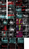
Regulation of dMyc expression by Notch and Wg. (AN, P) Wild type (A, B), Notchts2 reared at the restrictive temperature for 48 h (C), apGal4, UASGFP; UASmamDN (D), C96Gal4; UASmamDN (E, H), C96Gal4; UASTCFDN (F), C96Gal4; UASTCFDNUASNintra (G), C96Gal4; UASmamDN UASdMycdsRNA (I), dppGal4; UASNintra (J, K), apGal4, Gal80ts, UASGFP; UASTCFDN (L, N) dppGal4; UASWg (M), and apGal4, Gal80ts, UASGFP; UASTCFDN, UASmamDN (P) late third instar wing discs labelled to visualize dMyc protein (blue or white (A, D, E–J, M, N, P)) or dMyc mRNA (B, C, K, L) expression, and Wingless (Wg, red (A, E, J, M)), Cut (red (F, G)), Distalless (Dll, red (N, P)) or GFP (green (D, N, P)) protein expression. The DV compartment boundary in panels A and B is indicated by a dashed line. Note dMyc expression in GFP-expressing cells abutting the DV compartment boundary in D (compare with A) as well as in ZNC cells lacking Notch activity (C, E, H). (O, Q, R) ap–Gal4, Gal80ts, UAS–GFP; UAS–TCFDN, UAS–dMyc (O), dpp–Gal4; UAS–Nintra, UAS–dMyc (Q, R) late third instar wing discs labelled to visualize cells in S-phase by BrdU incorporation (antibody to BrdU in red or white (O, Q)), or expressing the E2F1-responsive reporter OCT1–GFP (pink (R)). GFP (O) and dMyc (R) protein expression (green) is also shown. Arrowheads in panels Q and R indicate the dpp–gal4 expression stripe. Wing discs shown in panels L and N–P were reared at 29°C during 30 h. (S) Wild-type wing disc labelled to visualize dMyc (blue) and Distalless (Dll, red) protein expression.
Figure 6
Figure 6
Regulation of bantam activity by Notch and Wg. (AG; I, J) Wild type (A, A′, E), apGal4; UASmamDN (B), dppGal4; UASNintra (C, C′), dpp–Gal4; UASWg (D), C96Gal4; UASTCFDN (F), C96Gal4; UASTCFDNUASNINTRA (G), dppGal4; UASTCFDN (I) and dppGal4; UASTCFDN, UASmamDN (J) late third instar wing discs labelled to visualize expression of the bantam sensor (blue and white), and BrdU incorporation (red (A)), Cut (red (A′)), Gal4 (red (B)), Wg (red (C, C′, I)) or Sens (red (D–G, J)) protein expression. Note symmetric expression of the bantam sensor in dorsal and ventral cells abutting the DV compartment boundary (dashed line) in panels A and A′, and lack of expression of the bantam sensor in dorsal cells abutting the DV compartment boundary (dashed line) in panel B (see also magnification). Note elevated levels of expression of the bantam sensor in the stripe of dppGal4 expression (red arrowheads) in panels C and I, when compared with panels A, B, D and J. (H, KM) C96Gal4; UASTCFDN/bantamΔ1 (H), dppGal4; UASTCFDN, UASbanGFP (K), dpp–Gal4; UAS–Nintra, UAS–ban–GFP (L) and dpp–Gal4; UAS–ban (M) late third instar wing discs labelled to visualize BrdU incorporation (red and white H, K, L) or E2F activity (red or white, M). Cut (green, H), GFP (green, K, L), Sens (blue, K) and Wg (blue, L, M) protein expression is also shown. Wing discs shown in panels I–K were reared at 29°C during 30 h.
Figure 7
Figure 7
Crosstalk between dMyc and bantam. (A) Wild-type wing disc labelled to visualize the bantam sensor (blue) and dMyc protein expression (red and white). (B) ptcGal4; UASbantamGFP wing disc showing dMyc expression (red and white) and GFP (green). (C, D) Clones of cells lacking bantam activity (banΔ1) marked by the absence of GFP. dMyc protein expression is shown in red or white. Note dMyc expression in the clones (red arrows). (EG) salGal4; UASdMyc (E), dppGal4; UASNintra, UASdMyc (F), and salGal4; UASdMycdsRNA (G) wing discs labelled to visualize expression of the bantam sensor (blue) and dMyc protein (red in E, red or white in G). The DV compartment boundary is marked in panels E and F (d, dorsal; v, ventral). (H) ptcGal4; UASdMycdsRNA, UASbantamGFP late third instar wing disc labelled to visualize BrdU incorporation (red), GFP (green) and dMyc (blue or white) protein expression. (I) C96Gal4; UASbantam late third instar wing disc, grown at 18°C and labelled to visualize expression of the bantam sensor (blue), BrdU incorporation (red) and dMyc protein expression (white).
Figure 8
Figure 8
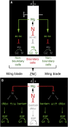
Different effects of Notch and Wg in cell-cycle control in the Drosophila wing. (A) Illustration describing the control of the G2-to-M transition in the anterior (a) compartment by the activity of Wg through its target genes achaete and scute (acsc). acsc are known to repress the expression of Drosophila cdc25 string (stg) in non-boundary cells. Note Wg does not exert any function in this process in posterior (p) cells. Notch represses acsc expression in boundary cerlls, this facilitating G2 progression in these cells. (B) Illustration describing the control of the G1–S transition by the activities of Notch (N) and Wingless (Wg) at the ZNC and in wing-blade cells, through the downstream effectors bantam and dMyc.

Similar articles

Cited by

References

    1. Asano M, Wharton RP (1999) E2F mediates developmental and cell cycle regulation of ORC1 in Drosophila. EMBO J 18: 2435–2448 - PMC - PubMed
    1. Axelrod JD, Matsuno K, Artavanis-Tsakonas S, Perrimon N (1996) Interaction between wingless and Notch signaling pathways mediated by Dishevelled. Science 271: 1826–1832 - PubMed
    1. Baena-Lopez LA, Garcia-Bellido A (2003) Genetic requirements of vestigial in the regulation of Drosophila wing development. Development 130: 197–208 - PubMed
    1. Baonza A, Freeman M (2005) Control of cell proliferation in the Drosophila eye by Notch signaling. Dev Cell 8: 529–539 - PubMed
    1. Bienz M, Clevers H (2000) Linking colorectal cancer to Wnt signaling. Cell 103: 311–320 - PubMed

Publication types

MeSH terms

LinkOut - more resources