. 2008 May 1;453(7191):115-9.
doi: 10.1038/nature06888.

Genome-wide screen reveals APC-associated RNAs enriched in cell protrusions

Affiliations

Genome-wide screen reveals APC-associated RNAs enriched in cell protrusions

Stavroula Mili et al. Nature. .

Abstract

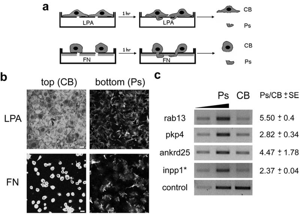
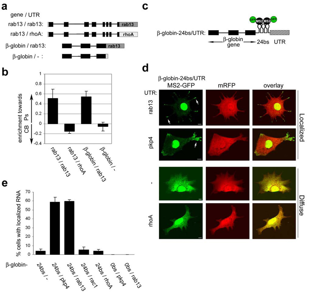
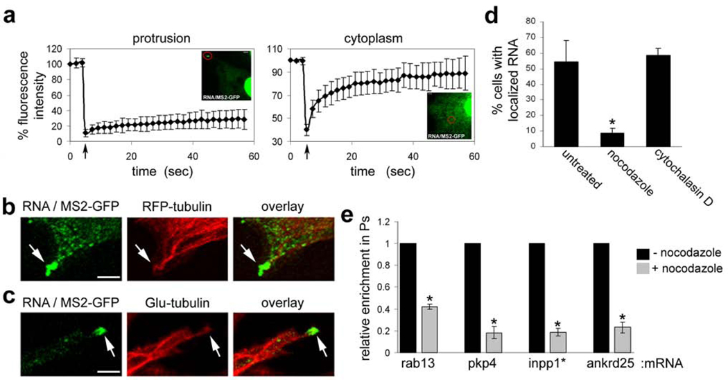
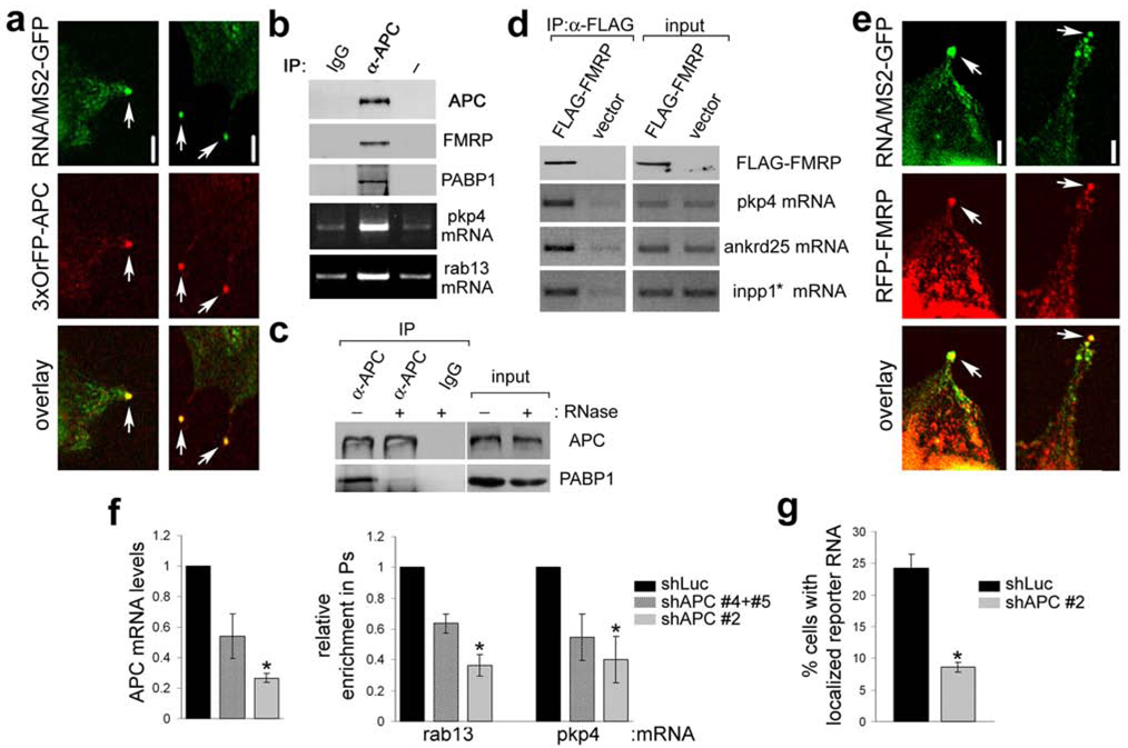
RNA localization is important for the establishment and maintenance of polarity in multiple cell types. Localized RNAs are usually transported along microtubules or actin filaments and become anchored at their destination to some underlying subcellular structure. Retention commonly involves actin or actin-associated proteins, although cytokeratin filaments and dynein anchor certain RNAs. RNA localization is important for diverse processes ranging from cell fate determination to synaptic plasticity; however, so far there have been few comprehensive studies of localized RNAs in mammalian cells. Here we have addressed this issue, focusing on migrating fibroblasts that polarize to form a leading edge and a tail in a process that involves asymmetric distribution of RNAs. We used a fractionation scheme combined with microarrays to identify, on a genome-wide scale, RNAs that localize in protruding pseudopodia of mouse fibroblasts in response to migratory stimuli. We find that a diverse group of RNAs accumulates in such pseudopodial protrusions. Through their 3' untranslated regions these transcripts are anchored in granules concentrated at the plus ends of detyrosinated microtubules. RNAs in the granules associate with the adenomatous polyposis coli (APC) tumour suppressor and the fragile X mental retardation protein (FMRP). APC is required for the accumulation of transcripts in protrusions. Our results suggest a new type of RNA anchoring mechanism as well as a new, unanticipated function for APC in localizing RNAs.


Figures

Figure 1
Figure 1. Several RNAs are enriched in protruding pseudopodia of migrating cells
a, Schematic depicting strategies used for isolation of pseudopodia (Ps) and cell bodies (CB). NIH/3T3 cells were allowed to attach onto FN-coated microporous filters and were induced to extend protrusions by adding LPA in the bottom chamber. Alternatively, only the underside of microporous filters was coated with FN and cells plated on top extended protrusions towards the FN-coated surface. CB and Ps fractions were physically isolated after 1 hr of induction. b, Cells extending protrusions in response to LPA or FN, as described in a, were fixed and stained with FITC-phalloidin. Confocal images of the top and bottom side of the filter are shown. Scale bar: 15µm. c, Total RNA was isolated from Ps and CB fractions of cells extending protrusions in response to LPA. The mRNAs indicated on the left were detected by quantitative RT-PCR. Increasing amounts of the Ps sample were amplified (lanes 1 and 2) to ensure linearity of the amplification. RNA levels were normalized to the control, arpc3, mRNA and the Ps/CB ratio was calculated. Values on the right indicate mean ± SEM of 3 independent experiments.
Figure 2
Figure 2. The 3’UTRs direct RNAs to accumulate in granules at tips of protrusions
a, Schematic of transfected constructs. Black boxes: exons, black lines: introns, grey boxes: 3’UTR, white box: FLAG tag. b, Cells transfected with the constructs depicted in a, were fractionated into Ps and CB fractions after induction with LPA. The enrichment of each RNA towards the Ps or CB fraction was calculated as described in Methods. Error bars: SEM c, Schematic depicting the general structure of β-globin constructs used in d, and e,. 24bs: 24 MS2-binding sites d, Cells were co-transfected with plasmids encoding mRFP, MS2-GFP and β-globin constructs with various 3’UTRs. Live cells were imaged during spreading on fibronectin. Shown are representative examples of the localization patterns observed when β-globin mRNA carried the UTRs indicated on the left; localized in granules at the tips of protrusions (arrows) or diffuse in the cytoplasm. Scale bar: 10µm e, Quantification of the percentage of cells exhibiting localized RNA distribution when transfected as in d, with constructs carrying the indicated 3’UTRs.
Figure 3
Figure 3. Localized RNA granules are anchored at the plus ends of detyrosinated microtubules
a, Cells were co-tranfected with constructs encoding MS2-GFP and the β-globin-24bs/pkp4 RNA. Fluorescence intensity in granules at protrusive areas (left panel, red circle) or within the cytoplasm (right panel, red circle) was monitored before and after photobleaching. Arrow indicates time of bleach. Curves represent average values of 10 and 5 independent experiments, respectively. Error bars: SD. b,c, Confocal fluorescence images of cells expressing MS2-GFP, the β-globin-24bs/pkp4 RNA and either RFP-tubulin (b) or fixed and stained with anti-Glu-tubulin antibody (c). Panels show edges of protrusive areas. Scale bar: 3µm. d, Cells expressing mRFP, MS2-GFP and the β-globin-24bs/pkp4 RNA were treated for 30 min with 10µM nocodazole or 1µM cytochalasin D. The percentage of cells exhibiting localized RNA distribution was quantified as in figures 2b and 2c. *: p-value<0.005 by two-tailed t-test versus untreated control. e, Cells on microporous filters were induced to extend protrusions in response to LPA in the absence or presence of 10µM nocodazole (added during the last 25 min of the assay). Enrichment of the indicated mRNAs in pseudopodia was determined as described in Methods and expressed relative to that of the untreated control cells. Values are averages of 3 independent experiments. Error bars: SEM. *: p-value<0.005 by paired, two-tailed t-test versus untreated control.
Figure 4
Figure 4. APC associates with RNP complexes containing FMRP and is required for localization of RNAs in protrusions
a, Confocal fluorescence images of cells expressing MS2-GFP, the β-globin-24bs/pkp4 RNA and 3xOrFP-APC. Panels show edges of protrusive areas. b, Lysates of NIH/3T3 cells were immunoprecipitated (IP) with control antibody (IgG), anti-APC antibody (α-APC) or no antibody (-) and analyzed to detect the indicated proteins and mRNAs. c, same as in b, except that prior to IP lysates were treated (+) or not (−) with RNase. d, Lysates of cells transfected with FLAG-FMRP or vector were immunoprecipitated with anti-FLAG antibody and analyzed to detect the indicated proteins and mRNAs. e, Confocal fluorescence images of cells expressing MS2-GFP, the β-globin-24bs/pkp4 RNA and RFP-FMRP. Panels show edges of protrusive areas. f, Cells expressing shRNAs against Luciferase (shLuc) or APC (shAPC), individually or in combination as indicated, were analyzed to determine APC mRNA levels. Shown are average values normalized to β-actin mRNA and expressed relative to that of the shLuc control (left panel). In parallel, cells were plated on filters and induced to extend protrusions in response to LPA. Enrichment of the indicated mRNAs in pseudopodia was determined as described in Methods and expressed relative to that of the shLuc control (right panel). Values are averages of 2–3 independent experiments. *: p-value<0.05 by paired, two-tailed t-test versus shLuc control. g, shLuc and shAPC-expressing cells were transfected with MS2-GFP and the β-globin-24bs/pkp4 RNA. The percentage of cells exhibiting localized RNA distribution, as described in Fig. 2d, was quantified. Values are averages of 7 independent experiments. *: p-value<0.0001 by two-tailed t-test. Error bars: SEM. Scale bar: 5µm.

References

    1. St Johnston D. Moving messages: the intracellular localization of mRNAs. Nat Rev Mol Cell Biol. 2005;6:363–375. - PubMed
    1. Forrest KM, Gavis ER. Live imaging of endogenous RNA reveals a diffusion and entrapment mechanism for nanos mRNA localization in Drosophila. Curr Biol. 2003;13:1159–1168. - PubMed
    1. Babu K, Cai Y, Bahri S, Yang X, Chia W. Roles of Bifocal, Homer, and F-actin in anchoring Oskar to the posterior cortex of Drosophila oocytes. Genes Dev. 2004;18:138–143. - PMC - PubMed
    1. Erdelyi M, Michon AM, Guichet A, Glotzer JB, Ephrussi A. Requirement for Drosophila cytoplasmic tropomyosin in oskar mRNA localization. Nature. 1995;377:524–527. - PubMed
    1. Jankovics F, Sinka R, Lukacsovich T, Erdelyi M. MOESIN crosslinks actin and cell membrane in Drosophila oocytes and is required for OSKAR anchoring. Curr Biol. 2002;12:2060–2065. - PubMed

Publication types

MeSH terms

Substances

Associated data