. 2008 Sep;15(9):1385-95.
doi: 10.1038/cdd.2008.59. Epub 2008 May 2.

BAC-mediated transgenic expression of fluorescent autophagic protein Beclin 1 reveals a role for Beclin 1 in lymphocyte development

Affiliations

BAC-mediated transgenic expression of fluorescent autophagic protein Beclin 1 reveals a role for Beclin 1 in lymphocyte development

I Arsov et al. Cell Death Differ. 2008 Sep.

Abstract

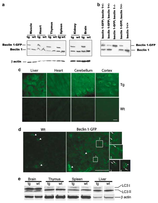
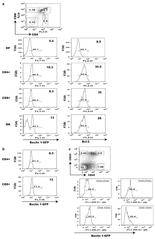
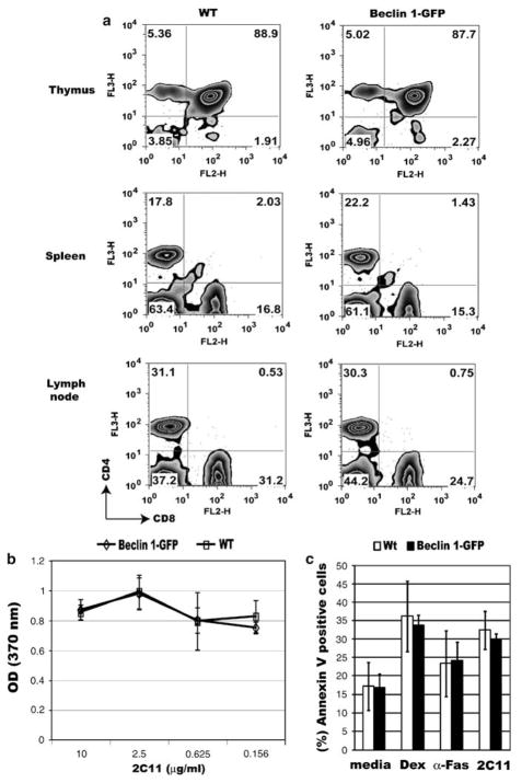
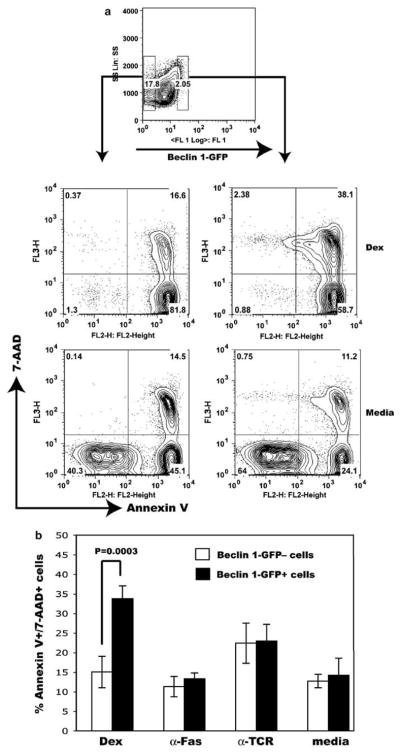
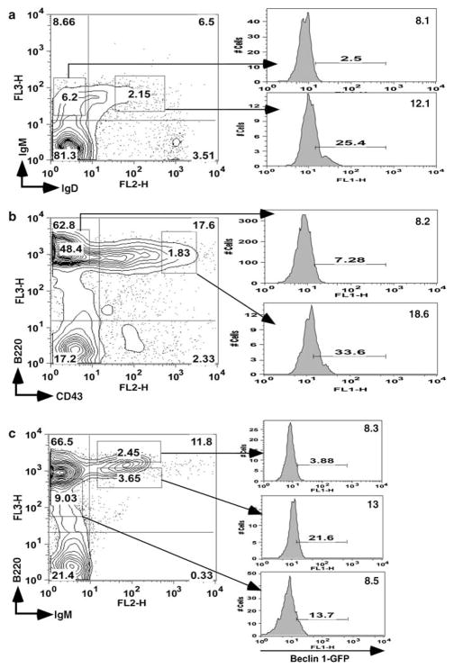
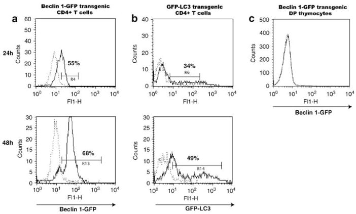
Beclin 1/Atg6 is an essential component of the evolutionary conserved PtdIns(3)-kinase (Vps34) protein complex that regulates macroautophagy (autophagy) in eukaryotic cells and also interacts with antiapoptotic Bcl-2 family members, Bcl-2, and Bcl-x(L). To elucidate the physiological function of Beclin 1, we generated transgenic mice producing a green fluorescent Beclin 1 protein (Beclin 1-GFP) under Beclin 1 endogenous regulation. The beclin 1-GFP transgene is functional because it completely rescues early embryonic lethality in beclin 1-deficient mice. The transgenic mice appear normal, with undetected change in basal autophagy levels in different tissues, despite the additional expression of functional Beclin 1-GFP. Staining of Beclin 1-GFP shows mostly diffuse cytoplasmic distribution in various tissues. Detailed analysis of the transgene expression by flow cytometry reveals a Bcl-2-like biphasic expression pattern in developing T and B cells, as well as differential regulation of expression in mature versus immature thymocytes following in vitro stimulation. Moreover, thymocytes expressing high Beclin 1-GFP levels appear increasingly sensitive to glucocorticoid-induced apoptosis in vitro. Our results, therefore, support a role for Beclin 1 in lymphocyte development involving cross talk between autophagy and apoptosis.


Figures

Figure 1
Figure 1
Expression levels and subcellular localization of functional Beclin 1-GFP protein in Beclin 1-GFP BAC transgenic mice. (a) Total protein extracts of various tissues from Beclin 1-GFP transgenic and control wild-type mice were analyzed with anti-Beclin 1 antibody and anti-β actin antibody. (b) Western blot analysis of Beclin 1 expression in muscle protein extracts, indicating the survival of beclin 1-GFP; beclin 1−/− mice. (c) Subcellular localization of Beclin 1-GFP analyzed by immunofluorescent staining with anti-GFP antibody reveals largely diffuse Beclin 1-GFP subcellular localization in the liver (hepatocytes), heart (cardiomyocytes) and cerebellum (Purkinje cells) or cortex (projection neurons) of the CNS. Top row, transgenic (Tg) tissues; bottom row, wild-type (Wt) tissues. (d) Immunofluorescent staining with anti-GFP antibody reveals a diffuse as well as punctate localization of Beclin 1-GFP in the thymus. Boxes 1 and 2 in the transgenic thymus are also shown in separate, enlarged pictures. Large arrows, autofluorescent dots; small arrows, Beclin 1-GFP positive puncta. Small fluorescent images 3 and 4 show live transgenic thymocytes in cell suspension. Scale bar, 20 μ. (e) Western blot analysis of endogenous LC3 (I and II) protein levels in the brain, thymus, spleen and liver of Wt mice and Tg mice.
Figure 2
Figure 2
Flow cytometry analysis of Beclin 1-GFP expression in major populations of transgenic T cells. (a) Beclin 1-GFP and Bcl-2 expression analysis in the thymus of Beclin 1-GFP transgenic mice. The percentage of Beclin 1-GFP positive cells within each thymocyte subset defined by corresponding gates in the top dot plot is indicated with small numbers within each histogram. The numbers in the upper right corner of each histogram represent mean fluorescence intensities (MFI) for each subset within indicated histogram gates. The histograms in (b) indicate Beclin 1-GFP expression in mature spleen CD4 + and CD8 + cells. (c) The histograms represent Beclin 1-GFP + fluorescence for each group of of pregated CD3CD4CD8 triple-negative Beclin 1-GFP + TN transgenic thymocytes defined by the gates indicated in the CD44 versus CD25 dot plot. Data are representative of at least four independent experiments
Figure 3
Figure 3
Normal T-cell development, activation, and apoptosis in Beclin 1-GFP transgenic mice. (a) Single-cell suspensions of the thymus, spleen, or lymph nodes from control (left panel), or Beclin 1-GFP transgenic mice (right panel) were stained with antibodies against CD4 and CD8 and analyzed on a FACScan flow cytometer. The percentage of gated cells in each quadrant is indicated. Data are representative of three independent experiments. (b) In vitro proliferation of purified Beclin 1-GFP transgenic (Tg) and control CD4 + T cells (Wt) stimulated with different concentrations of plate-bound 2C11 antibody and soluble anti-CD28 antibody (2 μg/ml) as described in Materials and Methods. (c) Apoptosis of Beclin 1-GFP transgenic and control thymocytes in vitro. Thymocytes from transgenic (Tg) and control (Wt) mice were incubated in the presence of either media alone (spontaneous apoptosis), dexamethasone (10−7 M), anti-Fas antibody (2 μg/ml), or precoated 2C11 antibody (10 μg/ml) for 24 h, cells stained with Annexin V and 7-AAD, and analyzed using flow cytometry. Late apoptotic/necrotic cells (Annexin V +7-AAD +) were excluded. Data represent means ± S.D. from three independent experiments
Figure 4
Figure 4
Accelerated dexamethasone-induced cell death in thymocytes expressing high Beclin 1-GFP levels. (a) Thymocytes expressing high levels of Beclin 1-GFP fluorescence, as well as Beclin 1-GFP-negative cells were sorted and cultured in vitro as described in Figure 4. The positions of corresponding gates were indicated with small rectangles (top panel). After 24 h in culture, cells were washed, stained with Annexin V and 7-AAD, and analyzed by flow cytometry. Only flow cytometry profiles obtained using cells cultured in dexamethasone and media from one representative experiment are shown. (b) Percent of late apoptotic/necrotic cells (Annexin V +7-AAD +) following stimulation of sorted transgenic thymocytes using indicated apoptotic stimuli. The results represent means ± S.D. from four independent experiments. Statistical analysis was performed using Student’s t-test. P < 0.05 was considered statistically significant
Figure 5
Figure 5
Analysis of Beclin 1-GFP expression in bone marrow B cells. Bone marrow cells from Beclin 1-GFP transgenic mice were stained with antibodies against (a) IgM and IgD, (b) CD43 and B220, and (c) IgM and B220, and analyzed by flow cytometry. The expression of Beclin 1-GFP in gated populations is shown in histograms on the right. Gate positions were indicated with rectangles. Numbers within histograms indicate percentage of GFP-positive cells using wild-type bone marrow as a negative control. Data are representative of three independent experiments
Figure 6
Figure 6
Stimulation of T cells upregulates Beclin 1-GFP and GFP-LC3 in CD4 + T cells. Purified (a) Beclin 1-GFP CD4 + T cells, (b) GFP-LC3 CD4 + T cells, or (c) Beclin 1-GFP total thymocytes were stimulated with plate-bound 2C11 antibody and soluble anti-CD28 antibody, as described in Materials and Methods (solid lines), or PBS (dotted lines) for 24 h (upper panels) or 48 h (lower panels), and analyzed for GFP expression using flow cytometry. The percentage of cells in each gate is indicated. Beclin 1-GFP fluorescence in (c) is shown for gated double-positive cells. Data are representative of three independent experiments

References

    1. Li C, Capan E, Zhao Y, Zhao J, Stolz D, Watkins SC, et al. Autophagy is induced in CD4+ T cells and important for the growth factor-withdrawal cell death. J Immunol. 2006;177:5163–5168. - PubMed
    1. Pua HH, Dzhagalov I, Chuck M, Mizushima N, He YW. A critical role for the autophagy gene Atg5 in T cell survival and proliferation. J Exp Med. 2007;204:25–31. - PMC - PubMed
    1. Klionsky DJ, Emr SD. Autophagy as a regulated pathway of cellular degradation. Science. 2000;290:1717–1721. - PMC - PubMed
    1. Levine B, Klionsky DJ. Development by self-digestion: molecular mechanisms and biological functions of autophagy. Dev Cell. 2004;6:463–477. - PubMed
    1. Mizushima N, Ohsumi Y, Yoshimori T. Autophagosome formation in mammalian cells. Cell Struct Funct. 2002;27:421–429. - PubMed

Publication types

MeSH terms