. 2008 May 2;4(5):e1000057.
doi: 10.1371/journal.ppat.1000057.

Primate lentiviral Vpx commandeers DDB1 to counteract a macrophage restriction

Affiliations

Primate lentiviral Vpx commandeers DDB1 to counteract a macrophage restriction

Natalia Sharova et al. PLoS Pathog. .

Abstract

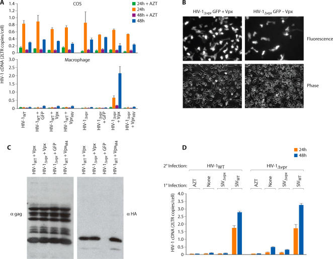
Primate lentiviruses encode four "accessory proteins" including Vif, Vpu, Nef, and Vpr/Vpx. Vif and Vpu counteract the antiviral effects of cellular restrictions to early and late steps in the viral replication cycle. We present evidence that the Vpx proteins of HIV-2/SIV(SM) promote virus infection by antagonizing an antiviral restriction in macrophages. Fusion of macrophages in which Vpx was essential for virus infection, with COS cells in which Vpx was dispensable for virus infection, generated heterokaryons that supported infection by wild-type SIV but not Vpx-deleted SIV. The restriction potently antagonized infection of macrophages by HIV-1, and expression of Vpx in macrophages in trans overcame the restriction to HIV-1 and SIV infection. Vpx was ubiquitylated and both ubiquitylation and the proteasome regulated the activity of Vpx. The ability of Vpx to counteract the restriction to HIV-1 and SIV infection was dependent upon the HIV-1 Vpr interacting protein, damaged DNA binding protein 1 (DDB1), and DDB1 partially substituted for Vpx when fused to Vpr. Our results indicate that macrophage harbor a potent antiviral restriction and that primate lentiviruses have evolved Vpx to counteract this restriction.


Conflict of interest statement

The authors have declared that no competing interests exist.

Figures

Figure 1
Figure 1. Vpx antagonizes an antiviral restriction in macrophages.
(A) Differential susceptibility of macrophages and cell lines to infection by wild type and Vpx-deleted (ΔVpx) or Vpr-deleted (ΔVpr) variants of SIVSM. Virus infection was gauged from the levels of viral 2-LTR cDNA at 24 and 48 h post infection. Right panels, comparison of infectivity of wild type and Vpx deleted SIV GFP variants. Macrophages were infected with 107, 106 or 105 RT unites of SIVWT -GFP or 107 RT units of SIVΔVpx GFP. Macrophages were visualized 48 h post infection by phase and fluorescence microscopy (B) Differential infectivity of wild type and Vpx-deleted SIV variants for macrophage-COS heterokaryons. Heterokaryons were formed between primary macrophages and between COS cells expressing fusogenic HN and F proteins of Newcastle Disease Virus. To visualize heterokaryons by fluorescence microscopy, COS cells were stained with DiO (green) and macrophages were stained with DiI (red) (magnification ×320; left panel). FACS analysis of macrophage-COS heterokaryons (middle panel). COS cells were cotransfected with NDV HN and F expression vectors (COS-NDV) or with empty, control vectors (COS). COS cells were stained with CellTracker Green CMFDA and macrophages were stained with CellTracker Blue CMAC. Double-stained cells were sorted as indicated by the gate. Susceptibility of macrophage/COS heterokaryons and COS and macrophage homokaryons to infection by SIVWT and SIVΔvpx virus variants (right panel). Infection was gauged from levels of late cDNAs and 2-LTR circle cDNAs (error bars are s.d. of 6 replicate samples from two independent experiments).
Figure 2
Figure 2. Vpx delivered to macrophages by SIVWT infection removes a block to subsequent infection by SIVΔvpx.
(A) Macrophages were initially infected (1° infection) with envelope deleted SIV variants harboring intact or defective Vpx genes. The nature of the envelope deletion is shown in the lower panel. Those cells were then super-infected (2° infection) with SIVWT or SIVΔvpx variants harboring intact envelope genes. cDNA products resulting from the super infection were then specifically amplified using envelope-specific primers. (B) Similar experiment was performed using SIVWT or SIVΔvpx for 1° infection and SIVWT or SIVΔvpx GFP variants for 2° infection. Number of GFP-positive cells was determined 24 hr post 2° infection (error bars are s.d., n = 3).
Figure 3
Figure 3. Role of the proteasome ubiquitylation system in regulation of SIV infectivity by Vpx.
(A) Identification of ubiquitylated residues in Vpx. Wild type and lysine mutant Vpx proteins (HA tag) were expressed in 293T cells expressing Histidine-tagged ubiquitin. (B) Susceptibility of primary macrophages to infection by SIVΔvpx packaging either wild type Vpx or lysine substitution mutants of Vpx. Vpx proteins were packaged after co-transfection of SIVΔvpx proviral DNA with plasmids expressing wild or lysine mutant Vpx proteins or GFP as a control. Virus infection was gauged from quantitation of late viral cDNAs and 2-LTR cDNAs 48 h post infection (error bars are s.d. of 3 replicate measures of a single DNA sample). (C) Packaging of wild type and non-ubiquitylated Vpx proteins in virus particles (upper panel). The presence of Vpx in gradient purified virions was determined by Western blotting with an HA antibody (lower panel).
Figure 4
Figure 4. Differential impact of proteasome inhibition on SIVWT and HIV-1 infection of macrophages.
Effects of 3 different proteasome inhibitors on SIV infection of macrophages and COS cells and HIV-1 infection of macrophages are indicated. Viral infection (2-LTR cDNA) was gauged at 24 and 48 h post infection (error bars and s.d. of 3 replicate measures of a single DNA sample).
Figure 5
Figure 5. HIV-1 is sensitive to the macrophage restriction and SIV Vpx but not HIV-1 Vpr antagonizes the restriction.
(A) HIV-1 Vpr and SIV Vpx proteins were packaged in HIV-1WT or HIV-1Δvpr viruses by cotransfection (for controls, viruses were transfected with an empty vector or a GFP-expressing vector). The infectivity of those viruses for COS (upper panel) and macrophages (lower panel) was then determined from levels of viral cDNA (2-LTR circle) at 24 and 48 h post infection. (B) Infection of macrophages with GFP-expressing HIV-1ΔVpr variants in which Vpx was (+) or was not (−) packaged. GFP positive macrophages (representative fields) were visualized 48 h post infection (C) Packaging of Vpx proteins in wild type and Vpr deleted HIV-1 (D) Vpx delivered to macrophages by SIVWT infection enhances the permissivity to HIV-1WT and HIV-1Δvpr infection. Macrophages were first infected (1° infection) with wild type or ΔVpx SIV variants, left uninfected (none) or treated with AZT. After 8 h, these cells were super-infected (2° infection) with WT or ΔVpr HIV-1 variants and HIV-1 infection (2-LTR cDNA synthesis) was determined 24 and 48 h later (error bars are s.d. of 3 replicate PCRs of a single DNA sample).
Figure 6
Figure 6. Ubiquitylation is required for enhancement of HIV-1 infectivity in macrophages.
(A) Infectivity of HIV-1 variants packaging wild type or lysine-mutant Vpx proteins. Wild type HIV-1 Vpr, SIV Vpx or lysine mutant Vpx (VpxM4) proteins were packaged in HIV-1Δvpr and infectivity of those viruses was assessed on macrophages. Viral cDNA synthesis was evaluated 24 and 48 h post-infection. (B) Infection of macrophages with HIV-1ΔVpr GFP variants packaging wild type or mutant Vpx proteins. Cells were visualized 48 h post-infection. (C) The ability of Vpx to enhance permissivity of macrophages to HIV-1 infection requires a functional proteasome. Macrophages were treated with the proteasome inhibitors ALLN or proteasome inhibitor 1 (Prot. 1) or with DMSO as a control. Those cells were then infected with HIV-1Δvpr variants which had packaged wild type or lysine mutant Vpx proteins. The level of viral infection (2-LTR cDNA) was determined 24 h post-infection by PCR (error bars are s.d. of 3 replicate PCRs of a single DNA sample.)
Figure 7
Figure 7. Inactivation of the macrophage restriction to SIV by Vpx requires DDB1.
(A) Association of SIV Vpx with endogenous DDB1. Association of DDB1 with wild-type Vpx (VpxWT) and non-ubiquitylated Vpx (VpxM4) was evaluated in 293T cells expressing FLAG-tagged Vpx (left panels) or HA-tagged Vpx (right panels) or IRES-GFP as a control. FLAG and HA immunoprecipitates were immunoblotted with DDB1 or FLAG and HA antibodies (upper panels). Levels of endogenous DDB1 and expressed Vpx in cell lysates were confirmed by Western blotting with a DDB1 antibody and with FLAG/HA antibodies respectively (lower panels). (B) Efficiency of siRNA-mediated silencing of DDB1 expression in COS cells and in macrophages was evaluated by Western blotting with DDB1 antibody at the indicated intervals post siRNA-transfection (ScrΙ-scrambled siRNA control). (C) Impact of DDB1 silencing on SIV and HIV-1 infection of COS cells and macrophages. SIV and HIV-1 infection was gauged from the quantity of viral cDNA (2-LTR) at 24, 48 and 72 h post infection (+, p>0.05; *, p<0.005) (error bars are s.d. of replicate PCRs of a single DNA sample).
Figure 8
Figure 8. DDB1 is required for the ability of Vpx to counteract the restriction to macrophage infection by HIV-1.
(A) SIV Vpx (or GFP as a control) was packaged into HIV-1ΔVpr virions as described in Figure 5. Infectivity of those viruses for DDB1-depleted macrophages (DDB1 siRNA) or control macrophages (ScrΙ siRNA) were evaluated from levels of viral cDNA 24 h later (+, p>0.2; *, p<0.002). (B) DDB1 packaging partially substitutes for Vpx. A Vpx/Vpr deletion mutant of SIV (SIVΔXR) was co-transfected with vectors expressing SIV Vpr, SIV Vpx, DDB1 or a Vpr-DDB1 fusion. Infectivity of the resulting viruses for macrophages was evaluated from levels of SIV cDNA at 24 and 48 h post infection (*, p<0.005). (C) Impact of DDB1 silencing on Vpx ubiquitylation. 293T cells were cotransfected with DDB1 or scrambled siRNAs and with HIS-ubiquitin and HA-Vpx or IRES-GFP expression plasmids as outlined in Figure 3. Ubiquitin-conjugated proteins were nickel purified and immunoblotted for Vpx (HA). Cell lysates were directly blotted for Vpx and DDB1 proteins (lower two panels).

References

    1. Emerman M, Malim MH. HIV-1 regulatory/accessory genes: keys to unraveling viral and host cell biology. Science. 1998;280:1880–4. - PubMed
    1. Sheehy AM, Gaddis NC, Choi JD, Malim MH. Isolation of a human gene that inhibits HIV-1 infection and is suppressed by the viral Vif protein. Nature. 2002;418:646–50. - PubMed
    1. Neil SJ, Zang T, Bieniasz PD. Tetherin inhibits retrovirus release and is antagonized by HIV-1 Vpu. Nature. 2008;24:406–8. - PubMed
    1. Fletcher TM, 3rd, Brichacek B, Sharova N, Newman MA, Stivahtis G, Sharp PM, Emerman M, Hahn BH, Stevenson M. Nuclear import and cell cycle arrest functions of the HIV-1 Vpr protein are encoded by two separate genes in HIV-2/SIMSM. EMBO J. 1966;15:6155–65. - PMC - PubMed
    1. Lu Y-L, Spearman P, Ratner L. Human immunodeficiency virus type 1 viral protein R localization in infected cells and virions. J Virol. 1993;67:6542–50. - PMC - PubMed

Publication types

MeSH terms

Substances