. 2008 Jun;118(6):2148-56.
doi: 10.1172/JCI33777.

Seven mutations in the human insulin gene linked to permanent neonatal/infancy-onset diabetes mellitus

Affiliations

Seven mutations in the human insulin gene linked to permanent neonatal/infancy-onset diabetes mellitus

Carlo Colombo et al. J Clin Invest. 2008 Jun.

Abstract

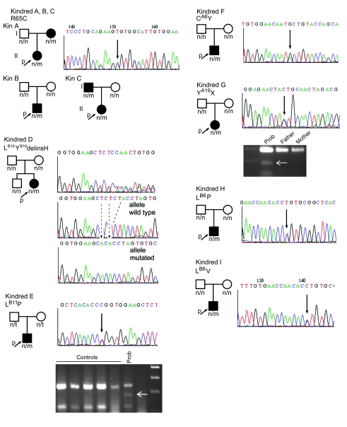
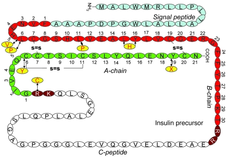
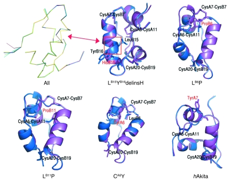
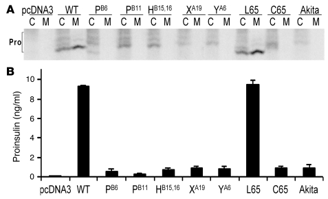
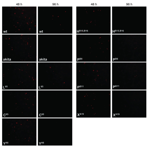
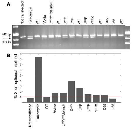
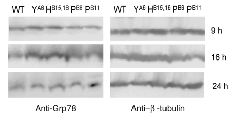
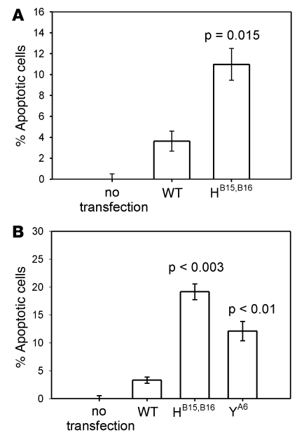
Permanent neonatal diabetes mellitus (PNDM) is a rare disorder usually presenting within 6 months of birth. Although several genes have been linked to this disorder, in almost half the cases documented in Italy, the genetic cause remains unknown. Because the Akita mouse bearing a mutation in the Ins2 gene exhibits PNDM associated with pancreatic beta cell apoptosis, we sequenced the human insulin gene in PNDM subjects with unidentified mutations. We discovered 7 heterozygous mutations in 10 unrelated probands. In 8 of these patients, insulin secretion was detectable at diabetes onset, but rapidly declined over time. When these mutant proinsulins were expressed in HEK293 cells, we observed defects in insulin protein folding and secretion. In these experiments, expression of the mutant proinsulins was also associated with increased Grp78 protein expression and XBP1 mRNA splicing, 2 markers of endoplasmic reticulum stress, and with increased apoptosis. Similarly transfected INS-1E insulinoma cells had diminished viability compared with those expressing WT proinsulin. In conclusion, we find that mutations in the insulin gene that promote proinsulin misfolding may cause PNDM.


Figures

Figure 1
Figure 1. Family trees of patients carrying INS gene mutations.
Pherograms of the mutations are also shown, with mutations indicated by black arrows. The mutation causing diabetes in kindred D, LB15YB16delinsH, was determined by subcloning of the PCR product and DNA sequencing, confirmed in 10 clones. Mutations LB11P and YA19X, which respectively introduce HpaII and SpeI restriction sites, were both confirmed (white arrows) by RFLP-PCR. n, normal allele; m, mutant allele; p, proband. Patients carrying mutations are denoted by filled symbols.
Figure 2
Figure 2. Amino acid sequences of WT and mutant proinsulins.
The amino acid sequence and disulfide bonds (s=s) of WT proinsulin are shown. Amino acid substitutions associated with PNDM/MDI are shown in yellow.
Figure 3
Figure 3. Secondary structure computer modeling of WT and mutant proinsulins.
Shown are superimposed Cα traces of WT (blue) and all mutated insulins (purple), with the exception of LB6V. The positions of disulfide bridges are also marked. None of the mutations caused substantial distortion of the secondary structure, with the exception of LB15YB16delinsH (yellow trace at left), where an α-helical disruption is apparent (arrow). This was also evident in the superimposition of WT insulin and LB15YB16delinsH. The LB6P mutation alters the hydrophobic core of the protein. The LB11P mutation affects the hydrophobic core of the protein. The CA6Y mutation disrupts the A6–A11 disulfide bridge; the tyrosine is oriented inside the hydrophobic core of the protein, where it engages LB6 in a stacking interaction. The hAkita mutation — used in this study as positive control — disrupts the B7–A7 disulfide bridge, and the tyrosine is solvent exposed.
Figure 4
Figure 4. Misfolding and defective secretion of proinsulin mutants.
293T cells were transfected with empty vector or cDNAs encoding WT proinsulin or the following proinsulin mutants: LB6P (PB6); LB11P (PB11); LB15YB16delinsH (HB15,16); YA19X (XA19); CA6Y (YA6); R65L (L65); R65C (C65); and hAkita. (A) Transfected cells were metabolically labeled with 35S–amino acids for 1 h and then further chased for 1 h. Cell lysates (C) and chase media (M) were immunoprecipitated with anti-insulin, and the samples were analyzed by nonreducing Tris-tricine-urea-SDS-PAGE. All proinsulin (Pro) disulfide isomers demonstrated different mobilities; the native form is the fast-migrating, secreted form obtained for WT proinsulin. (B) Recombinant proinsulin secreted for 16 h into serum-free medium was quantified by RIA.
Figure 5
Figure 5. Viability of transfected INS-1E cells.
Shown is the viability of INS-1E cells at 48 and 96 h after transfection with WT human (pro)insulin, hAkita mutation, human familial hyper(pro)insulinemia mutation R65L, and human MDI mutations R65C, CA6Y, LB15YB16delinsH, LB6P, LB11P, and YA19X. Human (pro)insulin was detected by monoclonal antibody directed toward the human C-peptide and C terminus of the B-chain of the (pro)insulin molecule. Few (LB6P and LB11P) or no (hAkita, R65C, CA6Y, LB15YB16delinsH, and YA19X) INS-1E cells expressing MDI mutations were visible 96 h after transfection.
Figure 6
Figure 6. XBP1 splicing in transfected HEK293 cells.
(A and B) Splicing of transcription factor XBP1 by agarose gel (A) and densitometry (B). Shown is the percentage of spliced/unspliced XBP1 forms for each sample, as assessed by densitometry, relative to WT. The red line indicates ratio 1. All mutations associated with PNDM or infancy-onset diabetes (and hAkita) induced an increase in XBP1 splicing compared with WT (pro)insulin or familial hyper(pro)insulinemia mutation R65L.
Figure 7
Figure 7. Grp78 expression.
Western blot with anti-Grp78 primary antibody in HEK293 cells transfected with WT and mutated insulin. Cytoplasmic fractions were collected at 9, 16, and 24 h after transfection. Expression of β-tubulin was assessed on the same filters to normalize the amount of loaded proteins. Increased Grp78 protein expression was clearly visible at 9 and 16 h for mutations LB15YB16delinsH and CA6Y and at 24 h for mutations LB6P and LB11P.
Figure 8
Figure 8. Annexin V expression and propidium iodide assay.
(A) Percentage of apoptotic cells, as assessed by annexin V expression, in HEK293 cells transfected with human WT (pro)insulin or LB15YB16delinsH mutation. Expression of mutant (pro)insulin effected a 3-fold increase in annexin V–positive cells 24 h after transfection compared with cells transfected with WT (pro)insulin. (B) Percentage of propidium iodide–positive cells transfected with WT, LB15YB16delinsH, and CA6Y mutation cDNA. P values are shown compared with WT (pro)insulin.

Similar articles

Cited by

References

    1. Polak M., Shield J. Neonatal and very-early-onset diabetes mellitus. Semin. Neonatol. 2004;9:59–65. doi: 10.1016/S1084-2756(03)00064-2. - DOI - PubMed
    1. Gloyn A.L., et al. Relapsing diabetes can result from moderately activating mutations in KCNJ11. Hum. Mol. Genet. 2005;14:925–934. doi: 10.1093/hmg/ddi086. - DOI - PubMed
    1. Colombo C., et al. Transient neonatal diabetes mellitus is associated with a recurrent (R201H) KCNJ11 (KIR6.2) mutation. . Diabetologia. 2005;48:2439–2441. doi: 10.1007/s00125-005-1958-1. - DOI - PubMed
    1. Babenko A.P., et al. Activating mutations in the ABCC8 gene in neonatal diabetes mellitus. . N. Engl. J. Med. 2006;355:456–466. doi: 10.1056/NEJMoa055068. - DOI - PubMed
    1. Stoffers D.A., Zinkin N.T., Stanojevic V., Clarke W.L., Habener J.F. Pancreatic agenesis attributable to a single nucleotide deletion in the human IPF1 gene coding sequence. Nat. Genet. 1997;15:106–110. doi: 10.1038/ng0197-106. - DOI - PubMed

Publication types

MeSH terms