. 2008 Jun;60(6):303-13.
doi: 10.1007/s00251-008-0296-0. Epub 2008 May 2.

Specificity of amyloid precursor-like protein 2 interactions with MHC class I molecules

Affiliations

Specificity of amyloid precursor-like protein 2 interactions with MHC class I molecules

Amit Tuli et al. Immunogenetics. 2008 Jun.

Abstract

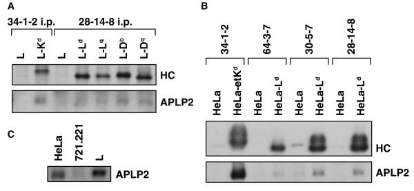
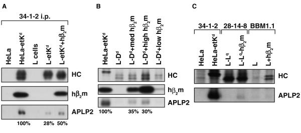
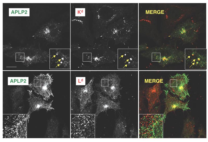
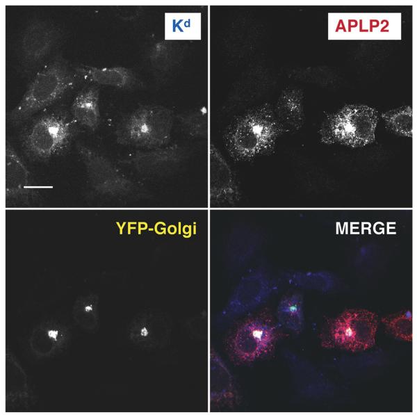
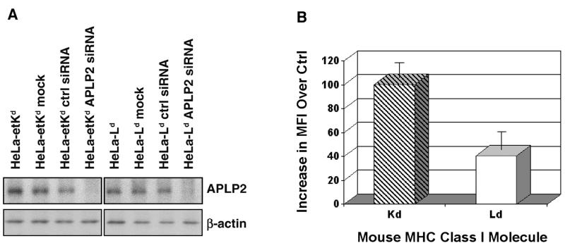
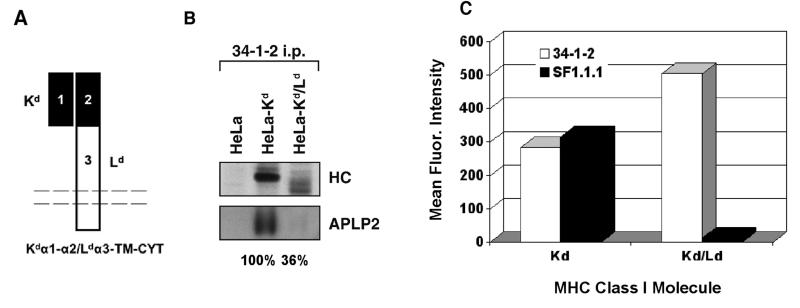
The ubiquitously expressed amyloid precursor-like protein 2 (APLP2) has been previously found to regulate cell surface expression of the major histocompatibility complex (MHC) class I molecule K(d) and bind strongly to K(d). In the study reported here, we demonstrated that APLP2 binds, in varied degrees, to several other mouse MHC class I allotypes and that the ability of APLP2 to affect cell surface expression of an MHC class I molecule is not limited to K(d). L(d), like K(d), was found associated with APLP2 in the Golgi, but K(d) was also associated with APLP2 within intracellular vesicular structures. We also investigated the effect of beta(2)m on APLP2/MHC interaction and found that human beta(2)m transfection increased the association of APLP2 with mouse MHC class I molecules, likely by affecting H2 class I heavy chain conformation. APLP2 was demonstrated to bind specifically to the conformation of L(d) having folded outer domains, consistent with our previous results with K(d) and indicating APLP2 interacts with the alpha1alpha2 region on each of these H2 class I molecules. Furthermore, we observed that binding to APLP2 involved the MHC alpha3/transmembrane/cytoplasmic region, suggesting that conserved as well as polymorphic regions of the H2 class I molecule may participate in interaction with APLP2. In summary, we demonstrated that APLP2's binding, co-localization pattern, and functional impact vary among H2 class I molecules and that APLP2/MHC association is influenced by multiple domains of the MHC class I heavy chain and by beta(2)m's effects on the conformation of the heavy chain.


Figures

Figure 1
Figure 1
(A) Kd, Db, and Dq expressed in L cells were weakly associated with APLP2. The indicated cell types were radiolabeled with 35 S-methionine/cysteine and immunoprecipitations were performed on lysates of the cells with 34-1-2 (for Kd) or 28-14-8 (for Ld, Lq, Db, or Dq). After electrophoresis on a 4→20% acrylamide gel, the immunoprecipitated proteins were transferred to a blotting membrane and autoradiographed to reveal the immunoprecipitated heavy chain (HC) or probed with antiserum recognizing APLP2 to reveal the co-immunoprecipitated APLP2. (B) Much more APLP2 was found associated with Kd than Ld in transfected HeLa cells. EtKd or Ld molecules expressed in HeLa cells were immunoprecipitated from cell lysates with 34-1-2 (for folded Kd), 64-3-7 (for open, peptide-free Ld), 30-5-7 (for folded Ld), or 28-14-8 (for both folded and open Ld). The immunoprecipitates were electrophoresed on 4-20% acrylamide gels and the proteins were transferred to a membrane and probed with 64-3-7 for the immunoprecipitated, denatured etKd or Ld MHC class I heavy chain (HC, top panel) or with antibody against APLP2 (bottom panel). Protein bands were quantified by multiplying the luminosity mean by the pixel number (as determined by Adobe Photoshop), and the ratio of the co-precipitated APLP2 band intensity to the Ld or Kd heavy chain band intensity was calculated. The results for the APLP2/30-5-7+ Ld ratio were 20% of the APLP2/34-1-2+ Kd ratio. (C) Expression levels of APLP2 in HeLa and L cells were demonstrated to be very similar. Samples of HeLa, 721.221, and L cell lysates were electrophoresed on a 4→20% gel, transferred to a blotting membrane, and probed with anti-APLP2 antiserum. Bands were quantified by multiplying the luminosity mean by the pixel number (as determined by Adobe Photoshop), and the ratio of the L cell APLP2 band to the HeLa APLP2 band was calculated to be 1.3.
Figure 2
Figure 2
(A)The presence of human β2m increased association of Kd with APLP2 in mouse L cells. Kd was immunoprecipitated from cell lysates with 34-1-2. The Kd immunoprecipitates were electrophoresed on 4→20% gels, and the proteins were transferred to blotting membranes. The membranes were probed with 64-3-7, which recognizes the Kd heavy chain (HC) denatured on the blot (top panel), with BBM1.1 that recognizes human β2m (middle panel), or with antiserum against APLP2 (bottom panel). Protein bands were quantified by multiplying the luminosity mean by the pixel number (as determined by Adobe Photoshop). The APLP2/etKd ratios for L-etKd and L-etKd+hβ2m are presented as percentages of the APLP2/Kd ratio for HeLa-etKd. (B) The presence of human β2m increased association of Dd with APLP2 in mouse L cells. Dd was immunoprecipitated from radiolabeled cell lysates with 34-1-2, electrophoresed on a 4→20% gel, and transferred to blotting membranes that were subsequently dried and autoradiographed to visualize the immunoprecipitated HC (top panel). The Dd immunoprecipitates were also electrophoresed on 4→20% gels for protein transfer to blotting membranes that were probed with BBM1.1 for human β2m (middle panel) or with antiserum against APLP2 (bottom panel). The APLP2/Dd ratios for L-Dd+med hβ2m and L-Dd+high hβ2m are presented as percentages of the APLP2/Kd ratio for HeLa-etKd. (C) APLP2 association was detectable for Lq after human β2m transfection into L-Lq, and β2m itself does not strongly associate with APLP2. The indicated immunoprecipitations from radiolabeled cell lysates were performed, and samples of the immunoprecipitates were electrophoresed on a 4→20% gel. The separated proteins were transferred to blotting membranes that were subsequently dried and autoradiographed to visualize the immunoprecipitated HC (top panel). The immunoprecipitates were also electrophoresed on 4→20% gels for protein transfer to blotting membranes that were probed with antiserum against APLP2 (bottom panel).
Figure 3
Figure 3
Both Kd and Ld were found to co-localize with APLP2 in a large intracellular compartment, but only Kd was found to co-localize with APLP2 in vesicles. (Top panel) At steady state, folded (34-1-2+) Kd was co-localized with APLP2. The co-localization was noted in a compact cellular compartment (identified as the Golgi in our earlier studies on Kd and APLP2) and also seen in peripheral vesicular structures. (Bottom panel) Folded (30-5-7+) Ld was co-localized with APLP2, but the co-localization could only be seen in the large compact compartment. For these experiments, HeLa-etKd or HeLa-Ld cells were transiently transfected with APLP2-FLAG, fixed, and incubated with 34-1-2 or 30-5-7 and rabbit anti-FLAG antibody and fluorescently labeled secondary antibodies in staining solution. Images were analyzed on a Zeiss LSM 5 Pascal confocal microscope. Green = APLP2; red = folded Kd or Ld; yellow = co-localized APLP2 and Kd or Ld molecules (single staining of APLP2 or Kd or Ld is shown in black and white in the figure). Bar, 10 μm.
Figure 4
Figure 4
The Golgi is the large compartment in which Kd and APLP2 co-localized. APLP2-FLAG, etKd, and YFP-Golgi were transiently transfected into HeLa cells. The cells were fixed and then incubated with anti-Kd antibody 34-1-2 and rabbit anti-FLAG and fluorescently labeled secondary antibodies in staining solution. Images were analyzed with a Zeiss LSM 5 Pascal confocal microscope. Blue = folded Kd; red=APLP2; yellow = YFP-Golgi; white = co-localized APLP2 and Kd (fluorescence from Kd, APLP2, and YFP-Golgi, individually, is shown in black and white in the figure). Bar, 10 μm.
Figure 5
Figure 5
Transient down-regulation of APLP2 slightly increased cell surface expression of Ld. (A) Samples from lysed HeLa-etKd or HeLa-Ld cells that had been untreated, mock treated, transfected with a control siRNA pool for 48 or 72 hours, or transfected with APLP2-specific siRNA for 48 or 72 hours were electrophoresed on a 4→20% gel, transferred to a blotting membrane, and probed with anti-APLP2 antiserum. Results from a 48 hour transfection are shown here, and results from 72 hours of transfection also showed down regulation (data not shown). (B) HeLa-etKd or HeLa-Ld cells that had been transfected with a control siRNA pool or with APLP2-specific siRNA for 48 or 72 hours were stained with 34-1-2 (for Kd) or 30-5-7 (for Ld) and a phycoerythrin-labeled secondary antibody, and then the fluorescence was assessed on a BD FACS Calibur. For each cell type, the mean fluorescence intensity (MFI) results in each of three experiments for cells expressing control siRNA were subtracted from the MFI results in the same experiment for cells transfected with APLP2-specific siRNA. The mean increases in MFI (i.e., for specific siRNA minus control siRNA) are shown in the graph with error bars indicating the standard error of the mean.
Figure 6
Figure 6
Substitution of the Ld α3/TM/CYT domain for the Kd α3/TM/CYT domain caused loss of APLP2 co-immunoprecipitation and increased cell surface expression. (A) A diagram of the Kd/Ld chimera. (B) Kd or Kd/Ld was immunoprecipitated from the indicated radiolabeled cell lysates with 34-1-2. The immunoprecipitates were electrophoresed on 4→20% gels, and the proteins were transferred to blotting membranes that were autoradiographed (HC, top panel) or probed with antiserum against APLP2 (bottom panel). (C) Shown on the bar graph are the mean fluorescence intensity values for Kd and Kd/Ld, as detected with antibody 34-1-2 (recognizing the Kd α1α2 domains) or SF1.1.1 (recognizing the Kd α3 domain) on Kd- or Kd/Ld-transfected HeLa cells. The MFI for Kd and Kd/Ld with fluorescently labeled secondary antibody only was less than 10 channels.

Similar articles

Cited by

References

    1. Achour A, Michaelsson J, Harris RA, Ljunggren H-G, Karre K, Schnieder G, Scandalova T. Structural basis of the differential stability and receptor-specificity of H-2Db in complex with murine versus human β2-microglobulin. J Mol Biol. 2006;356:382–396. - PubMed
    1. Benoit LA, Tan R. Xenogeneic β2-microglobulin substitution affects functional binding of MHC class I molecules by CD8+ T cells. J Immunol. 2007;179:3588–3595. - PubMed
    1. Brodsky FM, Bodmer WF, Parham P. Characterization of a monoclonal anti-beta 2-microglobulin antibody and its use in the genetic and biochemical analysis of major histocompatibility antigens. Eur J Immunol. 1979;9:536–545. - PubMed
    1. Cappai R, Mok SS, Galatis D, Tucker DF, Henry A, Beyreuther K, Small DH, Masters CL. Recombinant human amyloid precursor-like protein 2 (APLP2) expressed in the yeast Pichia pastoris can stimulate neurite outgrowth. FEBS Lett. 1999;442:95–98. - PubMed
    1. Cox JH, Bennick J, Yewdell JW. Retention of adenovirus E19 glycoprotein in the endoplasmic reticulum is essential to its ability to block antigen presentation. J Exp Med. 1991;174:1629–1637. - PMC - PubMed

Publication types

MeSH terms