

Characterization of a spontaneous, recessive, missense mutation arising in the Tecta gene
- PMID: 18452040
- PMCID: PMC2504605
- DOI: 10.1007/s10162-008-0116-0
Characterization of a spontaneous, recessive, missense mutation arising in the Tecta gene
Abstract
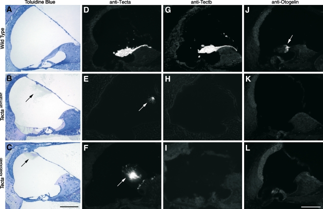
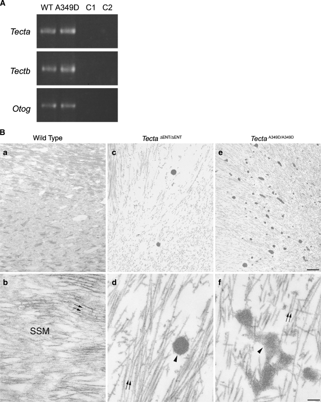
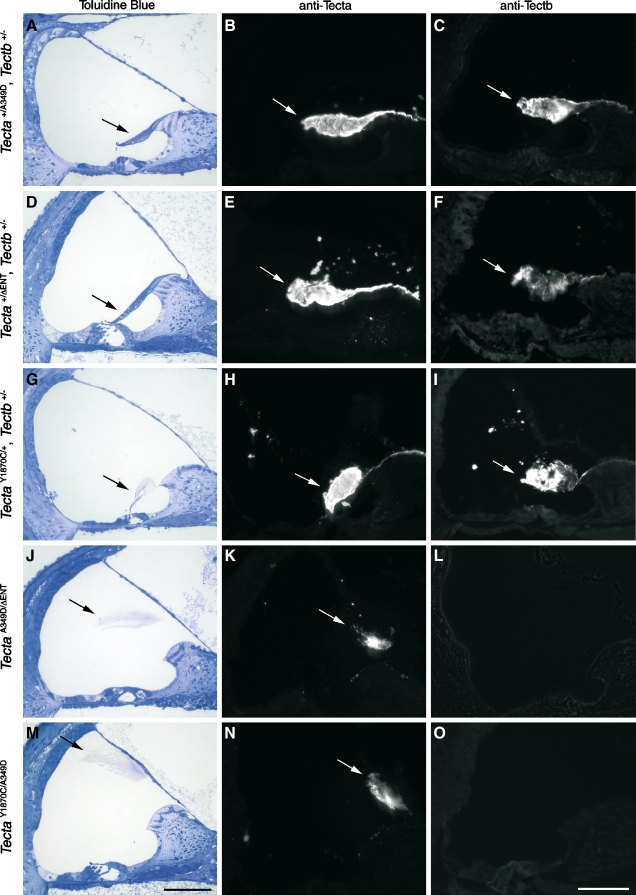
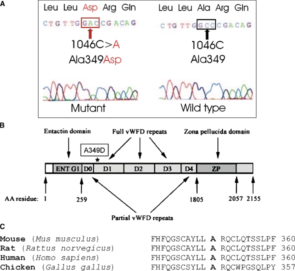
The TECTA gene encodes alpha-tectorin (TECTA), a major noncollagenous component of the tectorial membrane (TM). In humans, mutations in TECTA lead to either dominant (DFNA8/A12) or recessive (DFNB21) forms of nonsyndromic hearing loss. All missense mutations in TECTA that have been reported thus far are associated with the dominant subtype, whereas those leading to recessive deafness are all inactivating mutations. In this paper, we characterize a spontaneous missense mutation (c.1046C > A, p.A349D) arising in the mouse Tecta gene that is, unlike all previously reported missense mutations in TECTA, recessive. The morphological phenotype of the Tecta (A349D/A349D) mouse resembles but is not identical to that previously described for the Tecta(deltaENT)/(deltaENT) mouse. As in the Tecta(deltaENT/(deltaENT) mouse, the TM is completely detached from the surface of the organ of Corti and spiral limbus, lacks a striated-sheet matrix, and is deficient in both beta-tectorin (Tectb) and otogelin. A significant amount of Tecta is, however, detected in the TM of the Tecta (A349D/A349D) mouse, and numerous, electron-dense matrix granules are seen interspersed among the disorganized collagen fibrils. Mutated Tecta (A349D) is therefore incorporated into the TM but presumably unable to interact with either Tectb or otogelin. The Tecta (A349D/A349D) mouse reveals that missense mutations in Tecta can be recessive and lead to TM detachment and suggests that should similar mutations arise in the human population, they would likely cause deafness.
Figures
Similar articles
-
Identification of a Novel TECTA mutation in a Chinese DFNA8/12 family with prelingual progressive sensorineural hearing impairment.PLoS One. 2013 Jul 31;8(7):e70134. doi: 10.1371/journal.pone.0070134. Print 2013. PLoS One. 2013. PMID: 23936151 Free PMC article.
-
Three deaf mice: mouse models for TECTA-based human hereditary deafness reveal domain-specific structural phenotypes in the tectorial membrane.Hum Mol Genet. 2014 May 15;23(10):2551-68. doi: 10.1093/hmg/ddt646. Epub 2013 Dec 20. Hum Mol Genet. 2014. PMID: 24363064 Free PMC article.
-
An alpha-tectorin gene defect causes a newly identified autosomal recessive form of sensorineural pre-lingual non-syndromic deafness, DFNB21.Hum Mol Genet. 1999 Mar;8(3):409-12. doi: 10.1093/hmg/8.3.409. Hum Mol Genet. 1999. PMID: 9949200
-
[From gene to disease; DFNA8/12, an autosomal dominant inherited bowl-shaped sensorineural hearing impairment].Ned Tijdschr Geneeskd. 2007 Mar 3;151(9):531-4. Ned Tijdschr Geneeskd. 2007. PMID: 17373394 Review. Dutch.
-
The tectorial membrane: one slice of a complex cochlear sandwich.Curr Opin Otolaryngol Head Neck Surg. 2008 Oct;16(5):458-64. doi: 10.1097/MOO.0b013e32830e20c4. Curr Opin Otolaryngol Head Neck Surg. 2008. PMID: 18797289 Free PMC article. Review.
Cited by
-
Continued expression of GATA3 is necessary for cochlear neurosensory development.PLoS One. 2013 Apr 16;8(4):e62046. doi: 10.1371/journal.pone.0062046. Print 2013. PLoS One. 2013. PMID: 23614009 Free PMC article.
-
A novel splicing variant in TECTA associated with prelingual autosomal dominant nonsyndromic hearing loss via dominant-negative effect.Hum Mol Genet. 2025 Sep 3;34(18):1517-1525. doi: 10.1093/hmg/ddaf109. Hum Mol Genet. 2025. PMID: 40583560 Free PMC article.
-
Mutations of the gene encoding otogelin are a cause of autosomal-recessive nonsyndromic moderate hearing impairment.Am J Hum Genet. 2012 Nov 2;91(5):883-9. doi: 10.1016/j.ajhg.2012.09.012. Am J Hum Genet. 2012. PMID: 23122587 Free PMC article.
-
Mammalian Otolin: a multimeric glycoprotein specific to the inner ear that interacts with otoconial matrix protein Otoconin-90 and Cerebellin-1.PLoS One. 2010 Sep 15;5(9):e12765. doi: 10.1371/journal.pone.0012765. PLoS One. 2010. PMID: 20856818 Free PMC article.
-
Increased Spontaneous Otoacoustic Emissions in Mice with a Detached Tectorial Membrane.J Assoc Res Otolaryngol. 2016 Apr;17(2):81-8. doi: 10.1007/s10162-015-0551-7. Epub 2015 Dec 21. J Assoc Res Otolaryngol. 2016. PMID: 26691158 Free PMC article.
References
-
- Alasti F, Sanati MH, Behrouzifard AH, Sadeghi A, de Brouwer AP, Kremer H, Smith RJ, Van Camp G. A novel TECTA mutation confirms the recognizable phenotype among autosomal recessive hearing impairment families. Int. J. Pediatr. Otorhinolaryngol. 72:249–255, 2008. - PubMed
-
- {'text': '', 'ref_index': 1, 'ids': [{'type': 'DOI', 'value': '10.1038/sj.ejhg.5200273', 'is_inner': False, 'url': 'https://doi.org/10.1038/sj.ejhg.5200273'}, {'type': 'PubMed', 'value': '10196713', 'is_inner': True, 'url': 'https://pubmed.ncbi.nlm.nih.gov/10196713/'}]}
- Alloisio N, Morlé L, Bozon M, Godet J, Verhoeven K, Van Camp G, Plauchu H, Muller P, Collet L, Lina-Granade G. Mutation in the zonadhesin-like domain of α-tectorin associated with autosomal dominant non-syndromic hearing loss. Eur. J. Hum. Genet. 7:255–258, 1999. - PubMed
-
- {'text': '', 'ref_index': 1, 'ids': [{'type': 'DOI', 'value': '10.1007/s004390051091', 'is_inner': False, 'url': 'https://doi.org/10.1007/s004390051091'}, {'type': 'PubMed', 'value': '10987647', 'is_inner': True, 'url': 'https://pubmed.ncbi.nlm.nih.gov/10987647/'}]}
- Balciuniene J, Dahl N, Jalonen P, Verhoeven K, Van Camp G, Borg E, Pettersson U, Jazin E. Alpha-tectorin involvement in hearing disabilities: one gene-two phenotypes. Hum. Genet. 105:211–216, 1999. - PubMed
-
- {'text': '', 'ref_index': 1, 'ids': [{'type': 'PMC', 'value': 'PMC343085', 'is_inner': False, 'url': 'https://pmc.ncbi.nlm.nih.gov/articles/PMC343085/'}, {'type': 'PubMed', 'value': '987581', 'is_inner': True, 'url': 'https://pubmed.ncbi.nlm.nih.gov/987581/'}]}
- Blin N, Stafford DW. A general method for isolation of high molecular weight DNA from eukaryotes. Nucleic Acids Res. 3:2303–2308, 1976. - PMC - PubMed
-
- {'text': '', 'ref_index': 1, 'ids': [{'type': 'DOI', 'value': '10.1073/pnas.94.26.14450', 'is_inner': False, 'url': 'https://doi.org/10.1073/pnas.94.26.14450'}, {'type': 'PMC', 'value': 'PMC25017', 'is_inner': False, 'url': 'https://pmc.ncbi.nlm.nih.gov/articles/PMC25017/'}, {'type': 'PubMed', 'value': '9405633', 'is_inner': True, 'url': 'https://pubmed.ncbi.nlm.nih.gov/9405633/'}]}
- Cohen-Salmon M, El-Amraoui A, Leibovici M, Petit C. Otogelin: a glycoprotein specific to the acellular membranes of the inner ear. Proc. Natl. Acad. Sci. USA 94:14450–14455, 1997. - PMC - PubMed
Publication types
MeSH terms
Substances
Grants and funding
LinkOut - more resources
Full Text Sources
Research Materials
