

Review
doi: 10.1021/cr078215f.
Epub 2008 May 2.
The chemical neurobiology of carbohydrates
Affiliations
- PMID: 18452339
- PMCID: PMC4004190
- DOI: 10.1021/cr078215f
Item in Clipboard
Review
The chemical neurobiology of carbohydrates
Chem Rev.
2008 May.
No abstract available
Figures

Common structures of sialic acid derivatives: neuraminic acid (Neu), N-acetylneuraminic acid (Neu5Ac), N-glycolylneuraminic acid (Neu5Gc), and deaminoneuraminic acid (KDN).
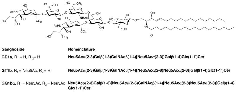
Structures of gangliosides that bind to MAG. Neu5Ac = N-acetylneuramic acid; Gal = galactose; GalNAc = N-acetylgalactosamine; Glc = glucose; Cer = ceramide.
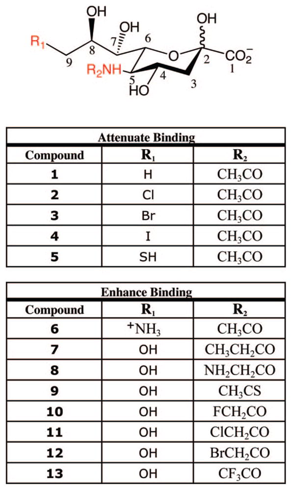
Synthetic sialic acid analogues tested for binding to MAG. Positions important for MAG interactions are shown in red.
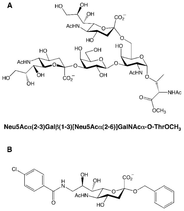
Structure of (A) a potent disialyl MAG inhibitor and (B) a simplified mimic of the ganglioside GQ1bα with enhanced binding affinity to MAG relative to Neu5Acα(2–3)Galβ(1–3)-GalNAc.

(A) Mannosamine derivatives used for metabolic labeling (R = H or Ac) and (B) chemoselective labeling reaction after treatment of cells with ManLev (R = biotin).

Structures of various fucose derivatives and 2-dGal.

Inhibition of Fucα(1–2)Gal linkages with 2-dGal leads to stunted neurite outgrowth in hippocampal neurons cultured for 4 days in vitro (DIV). D-Gal is able to rescue the effects of 2-dGal. 3-dGal has no effect. White bar indicates 45 μm. Images courtesy of C. Gama.
Chemical probe for imaging lectin receptors (top) and staining of hippocampal neurons in culture (bottom panels) with the probe demonstrating the presence of Fucα(1–2)Gal lectins along the cell body and neurites. Cells were treated with 3 mM of the imaging probe (A) or biotin (B), labeled with a streptavidin–dye conjugate, and imaged by fluorescence microscopy. Images courtesy of C. Gama.
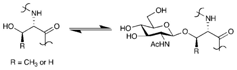
O-GlcNAc glycosylation.
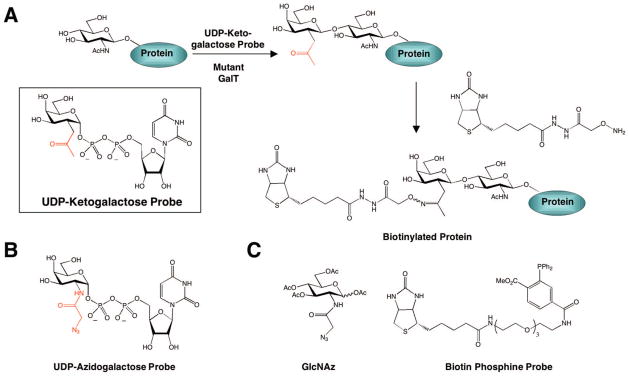
(A) Chemoenzymatic approach for tagging O-GlcNAc glycosylated proteins, (B) UDP–azidogalactose probe for [3 + 2] cycloaddition chemistry using the chemoenzymatic approach, and (C) GlcNAz and biotin phosphine probe for metabolic labeling of O-GlcNAc-modified protein using the Staudinger ligation.
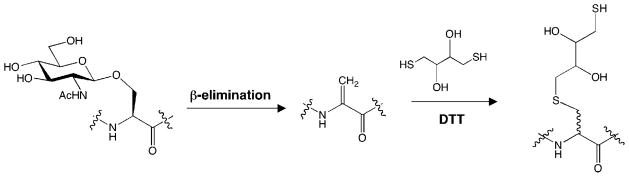
BEMAD approach for mapping O-GlcNAc glycosylation sites.
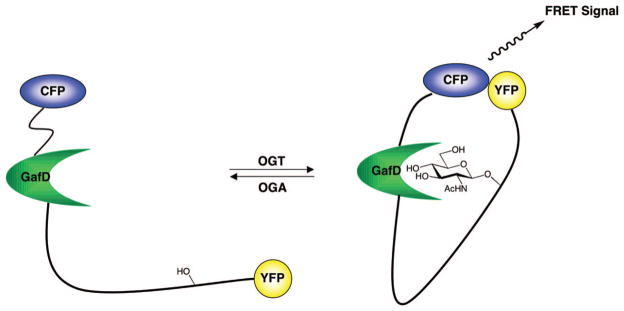
A fluorescence resonance energy transfer (FRET)-based sensor to detect O-GlcNAc glycosylation levels.

QUIC-Tag approach for quantifying dynamic changes in glycosylation.
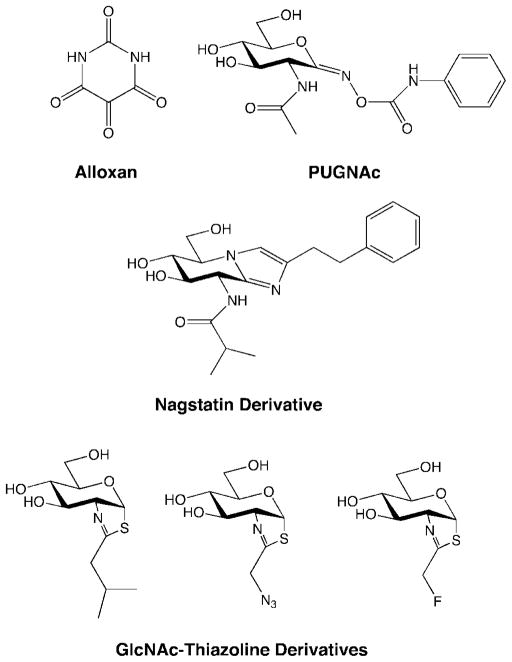
Small-molecule OGA inhibitors.

Structures of GAG subclasses. Potential sulfation sites are indicated in red. R = SO3− or H; R1 = SO3−, H, or Ac; n = ~10–200.

CS-E, -A, -C, and -R tetrasaccharides. Only the CS-E tetrasaccharide promotes neurite outgrowth.
References
-
- Becker DJ, Lowe JB. Glycobiology. 2003;13:41–53. - PubMed
-
- Gama CI, Hsieh-Wilson LC. Curr Opin Chem Biol. 2005;9:609–619. - PubMed
-
- Rampal R, Luther KB, Haltiwanger RS. Curr Mol Med. 2007;7:427–445. - PubMed
-
- Gabius HJ, Andre S, Kaltner H, Siebert HC. Biochim Biophys Acta. 2002;1572:165–177. - PubMed
-
- Nishihira J. Int J Mol Med. 1998;2:17–28. - PubMed
Publication types
MeSH terms
Substances
Grants and funding
LinkOut - more resources
Full Text Sources
Other Literature Sources
