. 2008 Jun;68(6):1485-501.
doi: 10.1111/j.1365-2958.2008.06247.x. Epub 2008 Apr 29.

YscU cleavage and the assembly of Yersinia type III secretion machine complexes

Affiliations

YscU cleavage and the assembly of Yersinia type III secretion machine complexes

Kelly E Riordan et al. Mol Microbiol. 2008 Jun.

Abstract

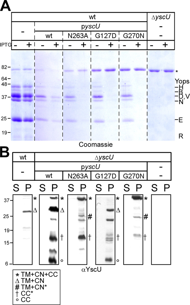
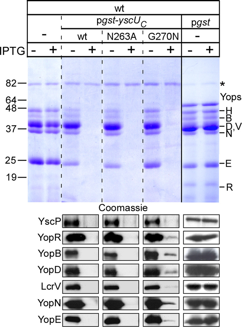
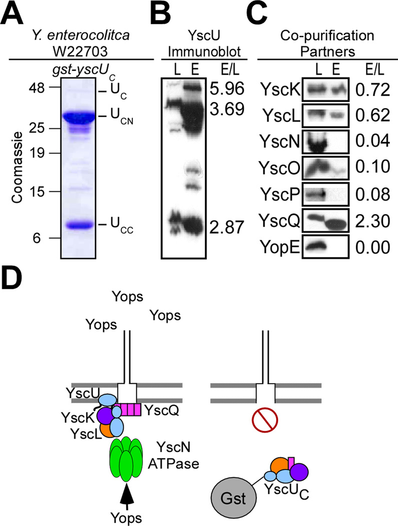
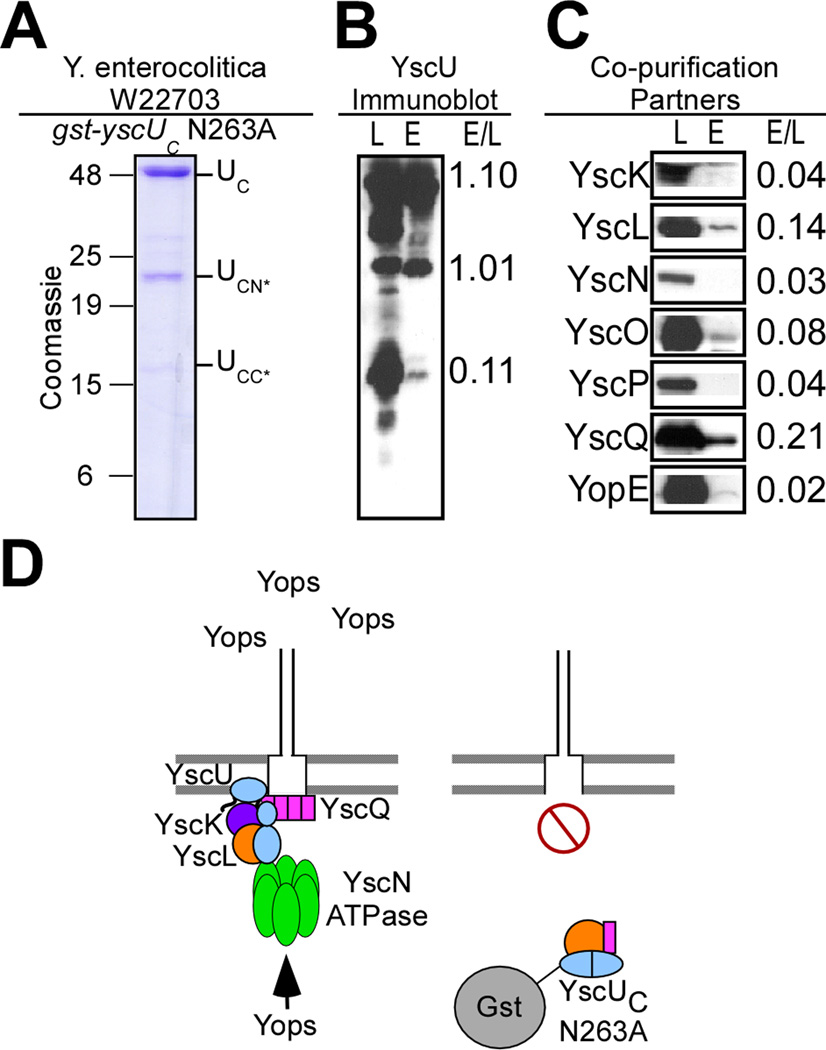
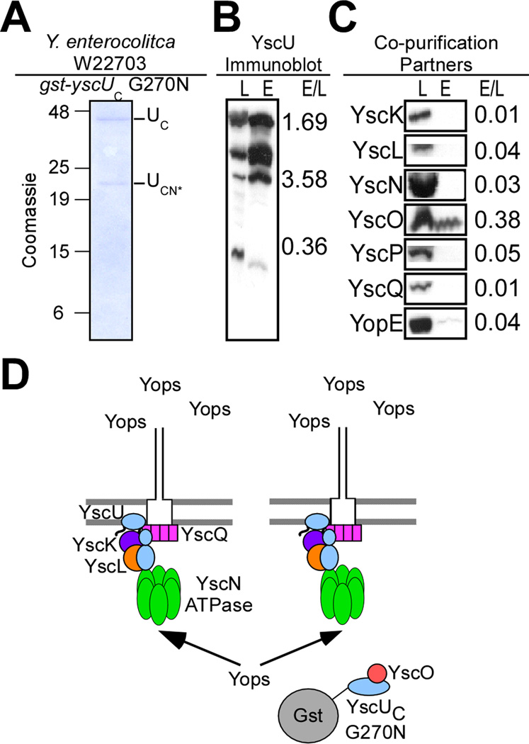
YscU, a component of the Yersinia type III secretion machine, promotes auto-cleavage at asparagine 263 (N263). Mutants with an alanine substitution at yscU codon 263 displayed secretion defects for some substrates (LcrV, YopB and YopD); however, transport of effector proteins into host cells (YopE, YopH, YopM) continued to occur. Two yscU mutations were isolated that, unlike N263A, completely abolished type III secretion; YscU(G127D) promoted auto-cleavage at N263, whereas YscU(G270N) did not. When fused to glutathione S-transferase (Gst), the YscU C-terminal cytoplasmic domain promoted auto-cleavage and Gst-YscU(C) also exerted a dominant-negative phenotype by blocking type III secretion. Gst-YscU(C/N263A) caused a similar blockade and Gst-YscU(C/G270N) reduced secretion. Gst-YscU(C) and Gst-YscU(C/N263A) bound YscL, the regulator of the ATPase YscN, whereas Gst-YscU(C/G270N) did not. When isolated from Yersinia, Gst-YscU(C) and Gst-YscU(C/N263A) associated with YscK-YscL-YscQ; however, Gst-YscU(C/G270N) interacted predominantly with the machine component YscO, but not with YscK-YscL-YscQ. A model is proposed whereby YscU auto-cleavage promotes interaction with YscL and recruitment of ATPase complexes that initiate type III secretion.


Figures

Fig. 1
Fig. 1
Phenotypic evaluation of hydroxylamine–generated mutants in the TTS gene yscU. (A) Y. pestis KIM D27 with chloramphenicol cassette inserted into the yopQ gene (MEL24) and ΔyscU (CHI110) Y. pestis containing vector control, pyscU (wt), pyscU N263A, pyscU G127D, and pyscU G270N on HIA plates containing 35 µg/mL chloramphenicol, 20 mM MgCl2, 20 mM oxalic acid, and 1 mM IPTG to induce type III secretion in the absence of calcium. Plates were grown at 37°C for 2 days to examine the LCR phenotype of indicated strains. (B) Diagram of the YscU protein indicating the four transmembrane helices and its hypothesized location in reference to the type III translocon. Mutations studied further are denoted with a red star. (C) Amino acid and corresponding DNA sequence of Y. enterocolitica W22703 yscU. Transmembrane domains are highlighted in light blue and the XNPTH cleavage motif highlighted in purple. All nonsense mutations obtained during hydroxylamine mutagenesis are denoted in green and missense mutations that generated phenotypes (G127D, G270N, N263A) are indicated in red. (D) Y. enterocolitica strains W22703, ΔyscU (CT-132), and ΔyscU containing pyscU (wt), pyscU N263A, pyscU G127D, and pyscU G270N were induced for type III secretion in TSB containing 35 µg/mL of chloramphenicol when needed for plasmid maintenance. Strains were grown in the absence of calcium (Ca2+) (5 mM EGTA) in the absence (−) or presence (+) of the inducer IPTG; and following centrifugation of culture aliquots, proteins in the supernatant (S) were separated by 12% SDS-PAGE. Gels were visualized with Coomassie blue staining; molecular mass markers (in kilodaltons) are indicated to the left of the gel while secreted Yop effectors are indicated to the right. An 80kD non-specific band common to all supernatants is indicated by an asterisk (*). (E) Supernatants generated in D were immunoblotted with polyclonal antisera specific to YopR, LcrV, YopB, YopD and YopE. The percent secretion of YopR, LcrV, YopB, YopD and YopE was calculated by comparing the secretion in each of the mutant strains to the amount of protein secreted in Y. enterocolitca ΔyscU containing pyscU (wt). Values are reported below the respective immunoblots. Secretion of YopR, LcrV, YopB, YopD, and YopE in Y. enterocolitca ΔyscU containing pyscU (wt) was set at 100%.
Fig. 2
Fig. 2
yscU N263A, but not G127D or G270N alleles support Y. enterocolitica type III injection into HeLa cells. (A) Y. enterocolitica strains W22703, ΔyscU (CT-132), and ΔyscU containing pyscU (wt), pyscU N263A, pyscU G127D, and pyscU G270N were used to infected HeLa tissue culture cells at an MOI of 10. IPTG was added to the medium to induce expression of various yscU alleles. The infection medium (M) was removed and centrifuged, separating the supernatant (S) and pellet (P). Digitonin was added to tissue culture cells with adherent bacteria. Digitonin extracted samples (D) were centrifuged, separating the supernatant (S) and pellet (P). All samples were precipitated with methanol/chloroform. Proteins were separated by 15% SDS-PAGE and immunoblotted with polyclonal antisera specific for YopR, YopE, YopH, YopN, YopB, YopD, LcrV, IκB, and RpoA. (B) HeLa tissue culture cells, 3 × 10⁵ cells/flask, were infected at an MOI of 10 with Y. enterocolitica strains W22703, ΔyscU (CT-132), and ΔyscU containing pyscU (wt), pyscU N263A, pyscU G127D, and pyscU G270N. Cytotoxicity of Y. enterocolitica infected HeLa cells was visualized by staining F-actin with rhodamine-conjugated phalloidin. Wild-type infected HeLa cells are shown as a control for type III injection.
Fig. 3
Fig. 3
Expression of pyscU N263A, G127D, and G270N in wild-type Y. enterocolitica shows dominant negative activity. (A) W22703 Y. enterocolitica strains expressing pyscU (wt), pyscU N263A, pyscU G127D, and pyscU G270N were induced for type III secretion (−Ca2+) in the absence (−) or presence (+) of IPTG. Following centrifugation of culture aliquots, proteins in the supernatant (S) were separated by 12% SDS-PAGE. Gels were visualized with Coomassie blue staining; molecular mass markers (in kilodaltons) are indicated to the left of the gel while secreted Yop effectors are indicated to the right. (B) Y. enterocolitica wild-type, ΔyscU containing pyscU (wt), pyscU N263A, pyscU G127D, and pyscU G270N, and ΔyscU were grown in the absence of calcium (Ca2+); and following centrifugation of culture aliquots, proteins in the supernatant (S) and bacterial pellet (P) were separated by SDS-PAGE and immunoblotted with polyclonal antisera specific to YscU (α-YscU). Several different species of the YscU protein were identified in the pellet fraction as denoted in the key.
Fig. 4
Fig. 4
Expression of wild-type Gst-YscUC and the N263A mutant produce dominant negative effects on type III secretion in Y. enterocolitica W22703. Y. enterocolitica W22703 containing plasmid-encoded gst-yscUC (pKER25), gst-yscUC N263A (pKER64), gst-yscUC (pKER68), and gst (pDA259) were induced for type III secretion in the absence (−) and presence (+) of IPTG. Following centrifugation of culture aliquots, proteins in the supernatant (S) were separated by 12% SDS-PAGE, analyzed by Coomassie blue staining, and immunoblotted with polyclonal antisera specific to YscP, YopR, YopB, YopD, LcrV, YopN, and YopE. Molecular mass markers (in kilodaltons) are indicated to the left of the gel and secreted Yop effectors are indicated to the right.
Fig. 5
Fig. 5
Gst-YscUC is cleaved at N263 and co-purifies in vivo with YscK, YscL, YscQ, and small amounts of YscO. (A) Y. enterocolitica W22703 expressing gst-yscUC (pKER25) under control of the tac promoter was induced for type III secretion in the absence of calcium. Cells were harvested by centrifugation and lysed via a French pressure cell. Samples were centrifuged at 100,000 ×g and separated into lysate supernatant (S) and pellet (P). The lysate supernatant (S) was subject to affinity chromatography on glutathione sepharose. A sample representing the eluate fraction was analyzed by 15% SDS-PAGE and Coomassie blue staining. Three major species in the elution (E) fractions at 42kD, 32kD, and 10kD were analyzed by mass spectrometry and identified as Gst-YscUC (aa 205–354) (UC), Gst-YscUCN (aa205–263) (UCN), and YscUCC (aa264–354) (UCC) respectively. (B) The lysate supernatant (L) and eluate fractions (E) were separated by SDS-PAGE and immunoblotted with antisera specific to YscU. Three species of YscU were detected via immunoblot. (C) For co-purification partners, the lysate supernatant (L) and eluate fractions (E) were separated by SDS-PAGE and immunoblotted with antisera specific to YscK, YscL, YscN, YscO, YscP, YscQ, and YopE. The relative intensity of immunoreactive signals in the lysate and eluate fractions (E/L) is expressed as a ratio to the right of each blot. (D) A model for the capture of machinery components and subsequent dominant negative effect of Gst-YscUC when expressed in wild-type Y. enterocolitica.
Fig. 6
Fig. 6
Purification of Gst-YscUC N263A reveals a unique cleavage product of 16kD (YscUCC*) and captures YscL, YscO, and YscQ, but not YscK in vivo. (A) Y. enterocolitica W22703 expressing gst-yscUC N263A (pKER64) under control of the tac promoter was induced for type III secretion in the absence of calcium. Cells were harvested by centrifugation and lysed via a French pressure cell. Samples were centrifuged at 100,000 ×g and separated into lysate supernatant (S) and pellet (P). The lysate supernatant (S) was subject to affinity chromatography on glutathione sepharose. A sample representing the eluate fraction was analyzed by 15% SDS-PAGE and Coomassie blue staining. Three major species in the elution (E) fractions at 42kD, 22kD, and 16kD were present and identified as Gst-YscUC (aa 205–354) (UC), Gst-YscUCN* (UCN*), and YscUCC* (UCC*) respectively. (B) The lysate supernatant (L) and eluate fractions (E) were separated by SDS-PAGE and immunoblotted with antisera specific to YscU. Two new species of YscU were detected via immunoblot. (C) The lysate supernatant (L) and eluate fractions (E) were separated by SDS-PAGE and immunoblotted with antisera specific to YscK, YscL, YscN, YscO, YscP, YscQ, and YopE. The relative intensity of immunoreactive signals in the lysate and eluate fractions (E/L) is expressed as a ratio to the right of each blot. (D) A model for the capture of machinery components and the subsequent dominant negative effect of Gst-YscUC N263A when expressed in wild-type Y. enterocolitica.
Fig. 7
Fig. 7
Gst-YscUC G270N co-purifies in vivo with YscO, but not YscK, YscL or YscQ. (A) Y. enterocolitica W22703 expressing gst-yscUC G270N (pKER68) under control of the tac promoter was induced for type III secretion in the absence of calcium. Cells were harvested by centrifugation and lysed via a French pressure cell. Samples were centrifuged at 100,000 ×g and separated into lysate supernatant (S) and pellet (P). The lysate supernatant (S) was subject to affinity chromatography on glutathione sepharose. A sample representing the eluate (E) fraction was collected and analyzed by 15% SDS-PAGE and Coomassie blue staining. Two major species in the elution (E) fractions at 42kD and 23kD were identified as Gst-YscUC (aa 205–354) (UC) and Gst-YscUCN* (UCN*). (B) The lysate supernatant (L) and eluate fractions (E) were separated by SDS-PAGE and immunoblotted with antisera specific to YscU. Three species of YscU were detected on immunoblot. (C) The lysate supernatant (L) and eluate fractions (E) were separated by SDS-PAGE and immunoblotted with antisera specific to YscK, YscL, YscN, YscO, YscP, YscQ, and YopE. The relative intensity of immunoreactive signals in the lysate and eluate fractions (E/L) is expressed as a ratio to the right of each blot. (D) A model for the capture of YscO by Gst-YscUC G270N when expressed in wild-type Y. enterocolitica.
Fig. 8
Fig. 8
Unlike Gst-YscUC wild-type and N263A, the G270N mutant does not capture His-YscL in vitro. (A) E. coli expressing His-yscK, His-yscL, His-yscN, His-yscQ, His-lcrV, and yopE-His were induced with 1 mM IPTG for expression of the fusion proteins. Cells were harvested by centrifugation and lysed via a French pressure cell. Samples were centrifuged at 100,000 ×g and separated into lysate supernatant (S) and pellet (P). Histidine tagged proteins were purified under denaturing conditions, separated by 15% SDS-PAGE and detected with Coomassie blue staining. Full length products are denoted by an arrow. The positions of molecular weight markers (in kilodaltons) are indicated to the left of the gel. (B) Wild-type, N263A, and G270N Gst-YscUC protein and Gst alone (10 µg charged to glutathione S-sepharose beads) were incubated with 10 µg each of His-YscK, His-YscL, His-YscN, His-YscQ, His-LcrV, and YopE-His. Samples were centrifuged and the supernatant precipitated with methanol-chloroform (unbound, U). Beads were washed three times with PBS and sedimented (bound, B). Both fractions were separated and analyzed by SDS-PAGE followed by immunoblot with polyclonal antisera to YscK, YscL, YscN, YscQ, LcrV, and YopE. Percent bound (%B) was calculated to be the relative intensity of immunoreactive signals in the bound fraction as compared to the total amount of immunoreactive protein (B/B+U) and is expressed as a percentage to the right of each blot.
Fig. 9
Fig. 9
A model for the role of YscU in the substrate specificity switch of Y. enterocolitica type III secretion machines. Uncleaved YscU is represented by two blue ovals signifying the YscUTM domain and uncleaved YscUC. Cleaved YscU is represented by three ovals signifying YscUTM, YscUCN, and YscUCC. It is proposed that YscO, binds to uncleaved YscUC allowing for secretion of early substrates, including the needle protein YscF, YscP, and YopR (YscH). Once needle assembly is completed and the needle sized by the YscP protein, YscU is signaled to undergo auto-cleavage at which point YscO dissociates from YscUC and may be secreted. Cleaved YscUCN+CC then recruits YscL to the base of the needle apparatus. YscL serves as the tether between YscUCN+CC and the YscK-YscQ-YscN ATPase complex.

Similar articles

Cited by

References

    1. Agrain C, Sorg I, Paroz C, Cornelis GR. Secretion of YscP from Yersinia enterocolitica is essential to control the length of the injectisome needle but not to change the type III secretion substrate specificity. Mol. Microbiol. 2005;57:1415–1427. - PubMed
    1. Aldridge P, Karlinsey JE, Becker E, Chevance FF, Hughes KT. Flk prevents premature secretion of the anti-sigma factor FlgM into the periplasm. Mol. Microbiol. 2006;60:630–643. - PMC - PubMed
    1. Allaoui A, Woestyn S, Sluiters C, Cornelis G. YscU, a Yersinia enterocolitica inner membrane protein involved in Yop secretion. J. Bacteriol. 1994;176:4534–4542. - PMC - PubMed
    1. Allaoui A, Schulte R, Cornelis GR. Mutational analysis of the Yersinia enterocolitica virC operon: characterization of yscE, F, G, I, J, K required for Yop secretion and yscH encoding YopR. Mol. Microbiol. 1995;18:343–355. - PubMed
    1. Anderson DM, Schneewind O. A mRNA signal for the type III secretion of Yop proteins by Yersinia enterocolitica. Science. 1997;278:1140–1143. - PubMed

Publication types