Review
. 2008 Jul;99(7):1319-25.
doi: 10.1111/j.1349-7006.2008.00840.x. Epub 2008 Apr 29.

Regulation of growth factor signaling by FRS2 family docking/scaffold adaptor proteins

Affiliations
Review

Regulation of growth factor signaling by FRS2 family docking/scaffold adaptor proteins

Noriko Gotoh. Cancer Sci. 2008 Jul.

Abstract

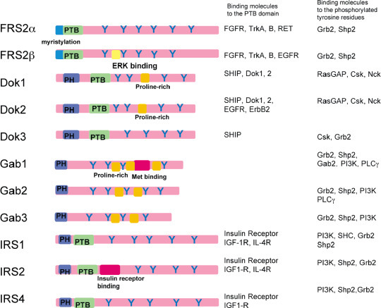
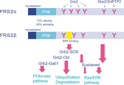
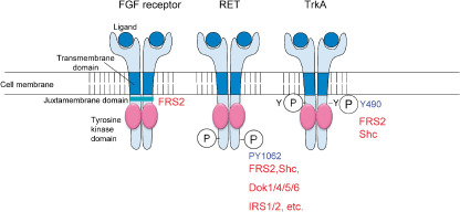
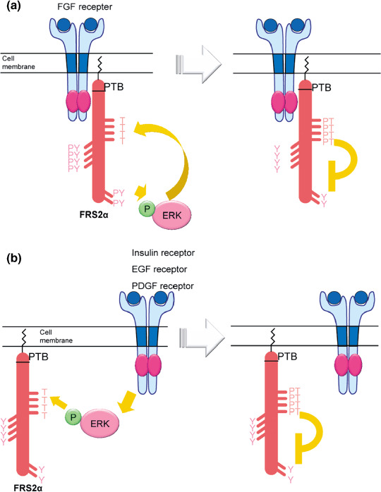
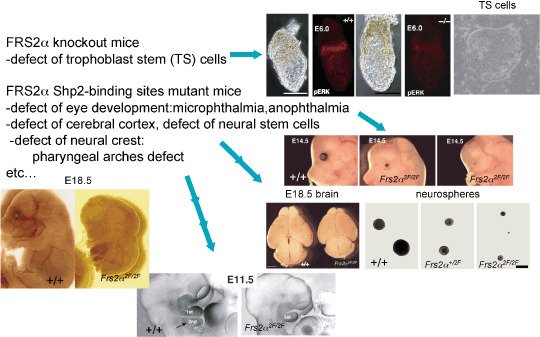
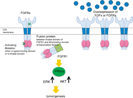
The FRS2 family of adaptor/scaffold proteins has two members, FRS2alpha and FRS2beta. Both proteins contain N-terminal myristylation sites for localization on the plasma membrane and a PTB domain for binding to limited species of receptor tyrosine kinases (RTKs), including the FGF receptor, the neurotophin receptor, RET, and ALK. Activation of these RTKs allows FRS2 proteins to become phosphorylated of tyrosine residues and then bind to Grb2 and Shp2, a SH2 domain-containing adaptor and a tyrosine phosphatase, respectively. Subsequently, Shp2 activates a Ras/ERK pathway and Grb2 activates a Ras/ERK, phosphatidyl inositol (PI)-3 kinase and ubiquitination/degradation pathways by binding to SOS, Gab1, and Cbl via the SH3 domains of Grb2. FRS2alpha acts as 'a conning center' in FGF signaling mainly because it induces sustained levels of activation of ERK via Shp2-binding sites and Grb2-binding sites, though the contribution of the former is greater. Indeed, FRS2alpha knockout mice and mice with mutated Shp2-binding sites exhibit a variety of phenotypes due to defects in FGF signaling in vivo. Although FRS2beta binds to the EGF receptor, it does not induce tyrosine phosphorylation on the receptor. Instead, it inhibits EGF signaling, resulting in inhibition of EGF-induced cell proliferation and cell transformation. Based on these findings, the involvement of FRS2 proteins in tumorigenesis should be studied extensively to be validated as candidate biomarkers for the effectiveness of treatments targeting RTKs such as the FGF receptor and EGF receptor.


Figures

Figure 1
Figure 1
Membrane‐linked docking proteins (MLDPs). Schematic structure of several MLDPs. The phosphotyrosine binding domain (PTB) binds to phosphrylated tyrosine residues on receptors or signaling proteins. The pleckstrin homology (PH) domain binds to phospholipids such as phosphatidyl inositol (PI)‐3 phosphate. The proline‐rich domains are important for binding to SH3 containing proteins such as Src family tyrosine kinases or Grb2. Y designates potential tyrosine (Y) phosphorylation sites. The ERK binding domain in FRS2β, Μet binding domain in Gab1, or insulin receptor binding domain in IRS2 contain a unique sequence for binding to ERK, Met, or insulin receptors.
Figure 2
Figure 2
Schematic structures of fibroblast growth factor (FGF) receptor substrate 2 (FRS2) family proteins. The phosphotyrosine binding domains (PTBs) of FRS2α and FRS2β have 72% identity and 93% similarity in amino acids. Tyrosine phoshporylated Grb2 binding sites activate Ras/ERK, ubiquination/degradation, and PI‐3 kinase pathways. Tyrosine phoshorylated Shp2 binding sites activate the Ras/ERK pathway stronger than the Grb2 binding site.
Figure 3
Figure 3
Binding sites of fibroblast growth factor (FGF) receptor substrate 2 (FRS2) proteins in receptor tyrosine kinases (RTKs). FRS2 binds to unphosphorylated peptides in the juxamembrane domain of the FGF receptor. It binds to tyrosine phosphorylated peptides in RET and TrkA, and the binding sites are shared with those of other PTB domain‐containing signaling proteins.
Figure 4
Figure 4
Negative feedback regulation of fibroblast growth factor (FGF) receptor substrate 2 (FRS2) proteins by activated ERK. (a) Activated FGF receptor phosphorylates tyrosine residues on FRS2α. The resultant activated ERK in turn phoshorylates threonine resiudes on FRS2α and inhibits tyrosine phosphorylation of FRS2α. (b) Activated insulin, epidermal growth factor (EGF), or the platelet‐derived growth factor (PDGF) receptor induce ERK activation without FRS2‐dependent mechanisms. The activated ERK phosphorylates threonine resiudes on FRS2α and inhibits tyrosine phosphorylation of FRS2α.
Figure 5
Figure 5
FRS2α is a ‘conning center’ in fibroblast growth factor (FGF) signaling in vivo. Frs2α null mouse embryos showed early embryonic lethality due to a failure of maintenance of trophoblast stem (TS) cells. TS cells are dependent on FGF4 and localized in extraembryonic ectoderm (ExE). The wild‐type ExE is positive in pERK staining but is weak in the mutant. The Frs2α 2F/2F mice have a variety of developmental defects, some of which are reasonably explained by the failure of FGF signaling.
Figure 6
Figure 6
Working hypothesis on the role of FRS2α in tumorigenesis. FRS2α appears to be involved in oncogenic addiction to aberrant fibroblast growth factor (FGF) signaling.

References

    1. Schlessinger J. Cell signaling by receptor tyrosine kinases. Cell 2000; 103: 211–25. - PubMed
    1. Pawson T, Scott JD. Signaling through scaffold, anchoring, and adaptor proteins. Science 1997; 278: 2075–80. - PubMed
    1. Pawson T. Dynamic control of signaling by modular adaptor proteins. Curr Opin Cell Biol 2007; 19: 112–16. - PubMed
    1. Nishida K, Hirano T. The role of Gab family scaffolding adapter proteins in the signal transduction of cytokine and growth factor receptors. Cancer Sci 2003; 94: 1029–33. - PMC - PubMed
    1. Gu H, Neel BG. The ‘Gab’ in signal transduction. Trends Cell Biol 2003; 13: 122–30. - PubMed

Publication types

MeSH terms

Substances