

Malignant gliomas with primitive neuroectodermal tumor-like components: a clinicopathologic and genetic study of 53 cases
- PMID: 18452568
- PMCID: PMC8094809
- DOI: 10.1111/j.1750-3639.2008.00167.x
Malignant gliomas with primitive neuroectodermal tumor-like components: a clinicopathologic and genetic study of 53 cases
Abstract
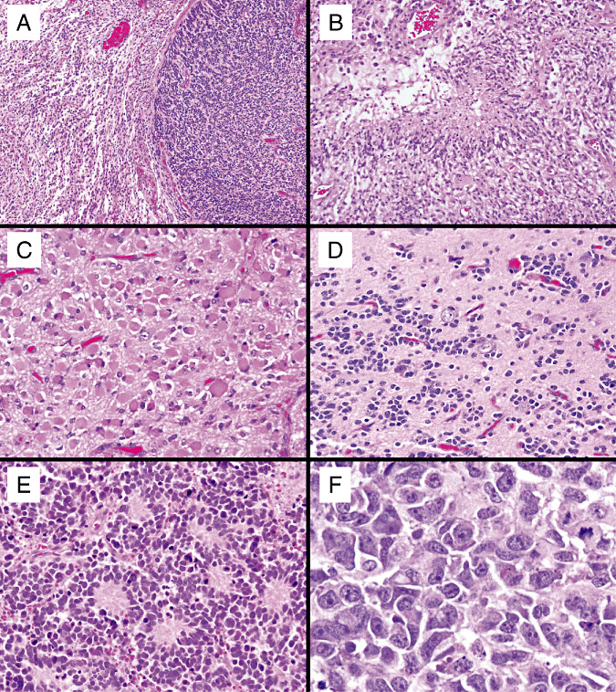
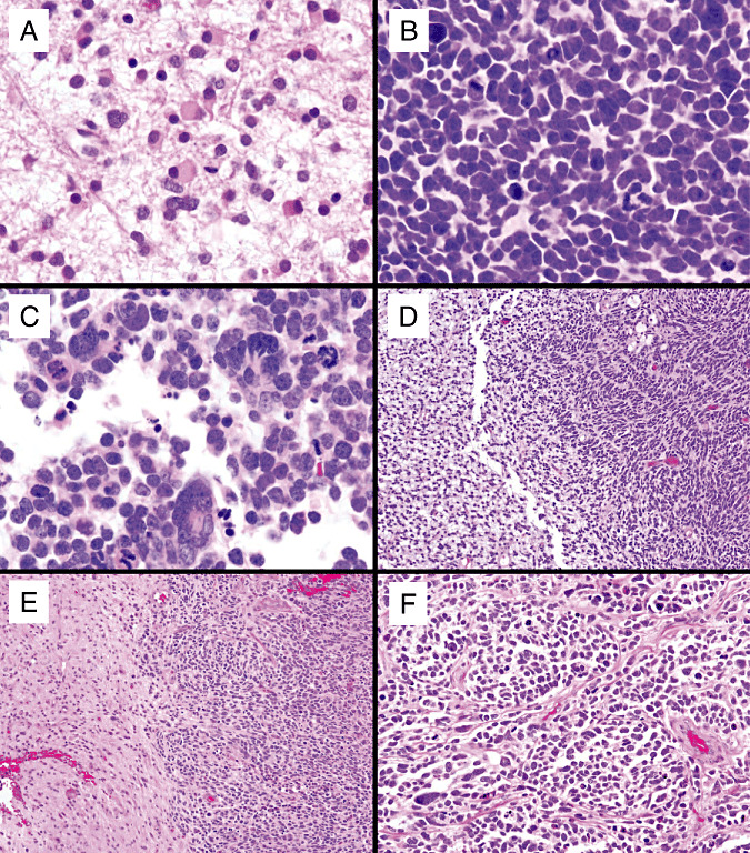
Central nervous system neoplasms with combined features of malignant glioma and primitive neuroectodermal tumor (MG-PNET) are rare, poorly characterized, and pose diagnostic as well as treatment dilemmas. We studied 53 MG-PNETs in patients from 12 to 80 years of age (median = 54 years). The PNET-like component consisted of sharply demarcated hypercellular nodules with evidence of neuronal differentiation. Anaplasia, as seen in medulloblastomas, was noted in 70%. Within the primitive element, N-myc or c-myc gene amplifications were seen in 43%. In contrast, glioma-associated alterations involved both components, 10q loss (50%) being most common. Therapy included radiation (78%), temozolomide (63%) and platinum-based chemotherapy (31%). Cerebrospinal fluid (CSF) dissemination developed in eight patients, with response to PNET-like therapy occurring in at least three. At last follow-up, 27 patients died, their median survival being 9.1 months. We conclude that the primitive component of the MG-PNET: (i) arises within a pre-existing MG, most often a secondary glioblastoma; (ii) may represent a metaplastic process or expansion of a tumor stem/progenitor cell clone; (iii) often shows histologic anaplasia and N-myc (or c-myc) amplification; (iv) has the capacity to seed the CSF; and (v) may respond to platinum-based chemotherapy regimens.
Figures


Similar articles
-
Malignant glioma with primitive neuroectodermal tumor-like component (MG-PNET): novel microarray findings in a pediatric patient.Clin Neuropathol. 2016 Nov/Dec;35(6):353-367. doi: 10.5414/NP300942. Clin Neuropathol. 2016. PMID: 27781423
-
Cytogenetic and histopathologic studies of congenital supratentorial primitive neuroectodermal tumors: a case report.Pathol Oncol Res. 2001;7(1):67-71. doi: 10.1007/BF03032609. Pathol Oncol Res. 2001. PMID: 11349224
-
Supratentorial primitive neuroectodermal tumors of the central nervous system in adults: molecular and histopathologic analysis of 12 cases.Am J Surg Pathol. 2011 Apr;35(4):573-82. doi: 10.1097/PAS.0b013e31820f1ce0. Am J Surg Pathol. 2011. PMID: 21378543
-
Primitive neuroectodermal tumors/medulloblastoma.Curr Neurol Neurosci Rep. 2002 May;2(3):205-9. doi: 10.1007/s11910-002-0078-2. Curr Neurol Neurosci Rep. 2002. PMID: 11936998 Review.
-
[CNS primitive neuroectodermal tumor suspected as a secondary recurrence after radiation therapy for medulloblastoma:a case report].No Shinkei Geka. 2014 Jul;42(7):641-50. No Shinkei Geka. 2014. PMID: 25006105 Review. Japanese.
Cited by
-
Leptomeningeal metastases in glioma: The Memorial Sloan Kettering Cancer Center experience.Neurology. 2019 May 21;92(21):e2483-e2491. doi: 10.1212/WNL.0000000000007529. Epub 2019 Apr 24. Neurology. 2019. PMID: 31019097 Free PMC article.
-
Glioblastomas with primitive neuronal component harbor a distinct methylation and copy-number profile with inactivation of TP53, PTEN, and RB1.Acta Neuropathol. 2021 Jul;142(1):179-189. doi: 10.1007/s00401-021-02302-6. Epub 2021 Apr 19. Acta Neuropathol. 2021. PMID: 33876327 Free PMC article.
-
Mitochondrial p32 is upregulated in Myc expressing brain cancers and mediates glutamine addiction.Oncotarget. 2015 Jan 20;6(2):1157-70. doi: 10.18632/oncotarget.2708. Oncotarget. 2015. PMID: 25528767 Free PMC article.
-
Paediatric and adult malignant glioma: close relatives or distant cousins?Nat Rev Clin Oncol. 2012 May 29;9(7):400-13. doi: 10.1038/nrclinonc.2012.87. Nat Rev Clin Oncol. 2012. PMID: 22641364 Review.
-
Maintenance of primary tumor phenotype and genotype in glioblastoma stem cells.Neuro Oncol. 2012 Feb;14(2):132-44. doi: 10.1093/neuonc/nor195. Epub 2011 Nov 7. Neuro Oncol. 2012. PMID: 22067563 Free PMC article.
References
-
- Actor B, Cobbers JM, Buschges R, Wolter M, Knobbe CB, Lichter P et al (2002) Comprehensive analysis of genomic alterations in gliosarcoma and its two tissue components. Genes Chromosomes Cancer 34:416–427. - PubMed
-
- Bailey P, Cushing H (1926) A Classification of Tumors of the Glioma Group on a Histogenetic Basis With a Correlation Study of Prognosis. Lippincott: Philadelphia, PA.
-
- Barbashina V, Salazar P, Ladanyi M, Rosenblum MK, Edgar MA (2007) Glioneuronal tumor with neuropil‐like islands (GTNI): a report of 8 cases with chromosome 1p/19q deletion analysis. Am J Surg Pathol 31:1196–1202. - PubMed
-
- Biernat W, Aguzzi A, Sure U, Grant JW, Kleihues P, Hegi ME (1995) Identical mutations of the p53 tumour suppressor gene in the glial and sarcomatous part of gliosarcomas suggest a common origin from glial cells. J Neuropathol Exp Neurol 54:651–656. - PubMed
-
- Boerman RH, Anderl K, Herath J et al (1996) The glial and mesenchymal elements of gliosarcomas share similar genetic alterations. J Neuropathol Exp Neurol 55:973–981. - PubMed
MeSH terms
Substances
LinkOut - more resources
Full Text Sources
Other Literature Sources
