. 2008 Jun;68(6):1595-613.
doi: 10.1111/j.1365-2958.2008.06265.x. Epub 2008 Apr 28.

Identification of a novel alpha(1-->6) mannopyranosyltransferase MptB from Corynebacterium glutamicum by deletion of a conserved gene, NCgl1505, affords a lipomannan- and lipoarabinomannan-deficient mutant

Affiliations

Identification of a novel alpha(1-->6) mannopyranosyltransferase MptB from Corynebacterium glutamicum by deletion of a conserved gene, NCgl1505, affords a lipomannan- and lipoarabinomannan-deficient mutant

Arun K Mishra et al. Mol Microbiol. 2008 Jun.

Abstract

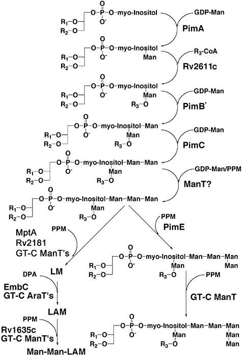
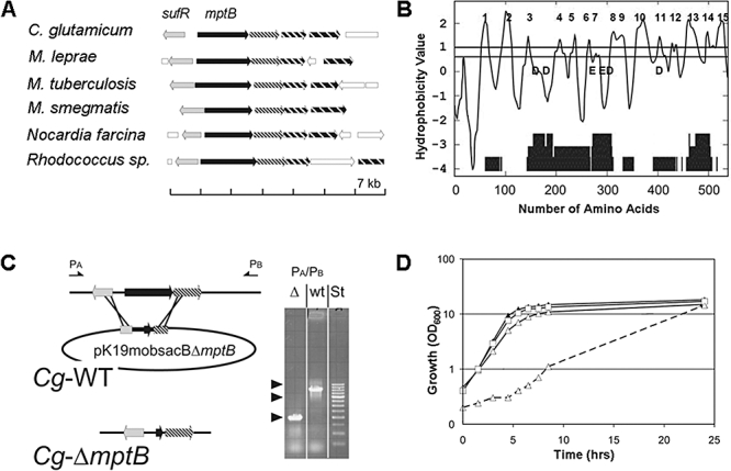
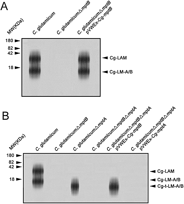
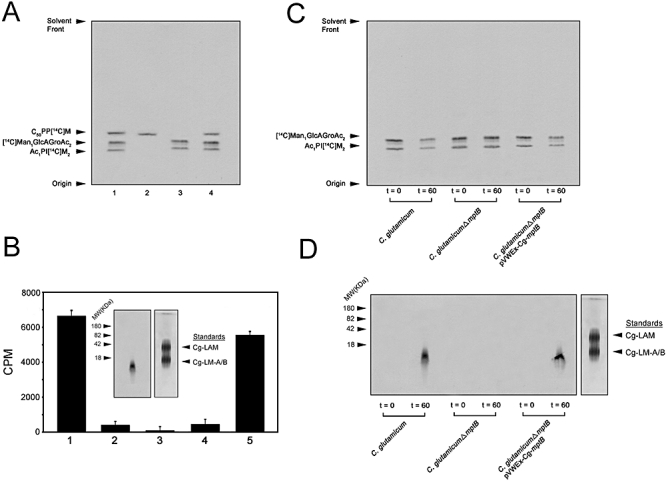
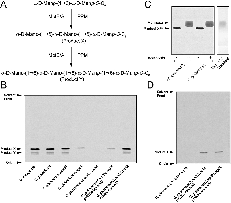
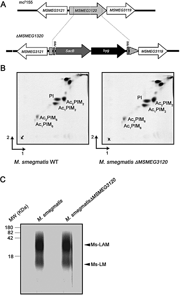
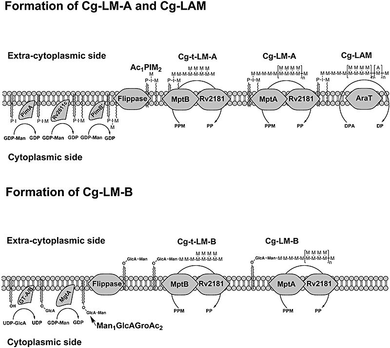
Mycobacterium tuberculosis and Corynebacterium glutamicum share a similar cell wall structure and orthologous enzymes involved in cell wall assembly. Herein, we have studied C. glutamicum NCgl1505, the orthologue of putative glycosyltransferases Rv1459c from M. tuberculosis and MSMEG3120 from Mycobacterium smegmatis. Deletion of NCgl1505 resulted in the absence of lipomannan (Cg-LM-A), lipoarabinomannan (Cg-LAM) and a multi-mannosylated polymer (Cg-LM-B) based on a 1,2-di-O-C(16)/C(18:1)-(alpha-D-glucopyranosyluronic acid)-(1-->3)-glycerol (GlcAGroAc(2)) anchor, while syntheses of triacylated-phosphatidyl-myo-inositol dimannoside (Ac(1)PIM(2)) and Man(1)GlcAGroAc(2) were still abundant in whole cells. Cell-free incubation of C. glutamicum membranes with GDP-[(14)C]Man established that C. glutamicum synthesized a novel alpha(1-->6)-linked linear form of Cg-LM-A and Cg-LM-B from Ac(1)PIM(2) and Man(1)GlcAGroAc(2) respectively. Furthermore, deletion of NCgl1505 also led to the absence of in vitro synthesized linear Cg-LM-A and Cg-LM-B, demonstrating that NCgl1505 was involved in core alpha(1-->6) mannan biosynthesis of Cg-LM-A and Cg-LM-B, extending Ac(1)PI[(14)C]M(2) and [(14)C]Man(1)GlcAGroAc(2) primers respectively. Use of the acceptor alpha-D-Manp-(1-->6)-alpha-D-Manp-O-C(8) in an in vitro cell-free assay confirmed NCgl1505 as an alpha(1-->6) mannopyranosyltransferase, now termed MptB. While Rv1459c and MSMEG3120 demonstrated similar in vitroalpha(1-->6) mannopyranosyltransferase activity, deletion of the Rv1459c homologue in M. smegmatis did not result in loss of mycobacterial LM/LAM, indicating a functional redundancy for this enzyme in mycobacteria.


Figures

Fig. 1
Fig. 1
Schematic representation of the current understanding of the LM and LAM biosynthetic pathway in M. tuberculosis. ManT, mannosyltransferase; AraT, arabinosyltransferase; PPM, polyprenyl-1-monophosphorylmannose; DPA, decaprenyl-1-monophosphoryl-arabinose; R1, R2 and R3 represent acyl groups.
Fig. 2
Fig. 2
Generation of an in-frame deletion mutant of C. glutamicum mptB. A. The locus in the bacteria analysed consists of mptB which has in C. glutamicum the locus tag NCgl1505 and in M. tuberculosis Rv1459c. sufR encodes a transcriptional regulator in front of an operon of the SUF machinery of [Fe-S] cluster synthesis (Huet et al., 2006). The genomic region displayed encompasses 7 kb, and orthologous genes are highlighted accordingly. Nocardia farcina, Nocardia farcina IFM 10152; Rhodococcus, Rhodococcus sp. strain RHA1. B. MptB is a hydrophobic protein predicted to span the membrane 15 times and the transmembrane helices are numbered accordingly. The lower part of the figure shows the degree of conservation of the orthologues given in A as analysed by the dialign method (Morgenstern, 2004). Also shown is the approximate position of the fully conserved aspartyl (D) and glutamyl (E) residues. C. Strategy to delete Cg-mptB using the deletion vector pK19mobsacBΔmptB. This vector carries 18 nucleotides of the 5′ end of Cg-mptB and 36 nucleotides of its 3′ end, thereby enabling the in-frame deletion of almost the entire Cg-mptB gene. The arrows marked PA and PB locate the primers used for the PCR analysis to confirm the absence of Cg-mptB. Distances are not drawn to scale. The results of the PCR analysis with the primer pair PA/PB are shown on the right. Amplification products obtained from the wild type (wt) were applied in the middle lane and that of the deletion mutant (Δ) in the left lane. ‘st’ marks the standard, where the arrowheads are located at 1.5, 1 and 0.5 kb. D. Growth of C. glutamicum on rich BHIS (solid lines). Wild-type C. glutamicm, filled triangle; C. glutamicumΔmptB, open triangle; C. glutamicumΔmptB pVWEx-Cg-mptB, open square. Growth of C. glutamicumΔmptB on rich BHI medium are the open triangles with the broken line.
Fig. 3
Fig. 3
Lipoglycan profile of C. glutamicum strains analysed using SDS-PAGE and visualized using a Pro-Q emerald glycoprotein stain (Invitrogen) specific for carbohydrates. A. Lipolglycans extracted from C. glutamicum, C. glutamicumΔmptB and C. glutamicumΔmptB pVWEx-Cg-mptB. The major bands represented by Cg-LAM, Cg-LM-A and Cg-LM-B are indicated. B. C. glutamicum, C. glutamicumΔmptB, C. glutamicumΔmptA, C. glutamicumΔmptBΔmptA, C. glutamicumΔmptBΔmptA pVWEx-Cg-mptB and C. glutamicumΔmptBΔmptA pVWEx-Cg-mptA. The truncated version of Cg-LM-A/B is indicated as Cg-t-LM-A/B (Mishra et al., 2007). The four major bands represent glycoproteins of 180, 82, 42 and 18 kDa respectively.
Fig. 4
Fig. 4
Incorporation of [14C]Man from GDP-[14C]Man into corynebacterial membrane/cell envelope lipids. A. TLC autoradiography of labelled CHCl3/CH3OH (2:1)-soluble lipids, C50-PP[14C]M, [14C]Man1GlcAGroAc2 and Ac1PI[14C]M2 using GDP-[14C]Man and membrane/cell envelope extracts from C. glutamicum and C. glutamicumΔmptB. Membrane/cell envelope fractions were incubated with GDP-[14C]Man in a total volume of 100 μl for 60 min in either the absence or presence of amphomycin (10 μg) and Ca2+ ions per reaction mixture pre-incubated with membranes for 15 min. Enzymatically synthesized products C50-PP[14C]M, [14C]ManGlcAGroAc2 and Ac1PI[14C]M2 were isolated as described in Experimental procedures to provide washed CHCl3/CH3OH (2:1)-soluble lipids and also subjected to base treatment. Aliquots (10%) were taken for scintillation counting and the remaining products subjected to TLC/autoradiography using CHCl3/CH3OH/NH4OH/H2O (65:25:0.4:3.6, v/v/v/v). C. glutamicum CHCl3/CH3OH (2:1)-soluble lipids (lane 1), base treatment of CHCl3/CH3OH (2:1)-soluble lipids (lane 2), amphomycin treatment (lane 3) and C. glutamicumΔmptB CHCl3/CH3OH (2:1)-soluble lipids (lane 4). B. Characterization of CHCl3/CH3OH/H2O (10:10:3)-soluble lipids as α(1→6)-linear mannooligosaccharides. The insoluble pellet from the above reaction mixtures following extraction with CHCl3/CH3OH (2:1) were sequentially washed with 0.9% NaCl in 50% CH3OH, 50% CH3OH and CH3OH, prior to extraction with CHCl3/CH3OH/H2O (10:10:3) and an aliquot (10%) taken for scintillation counting and the remaining product analysed by SDS-PAGE/autoradiography (left-panel inset). C. glutamicum (no. 1), amphomycin treatment (no. 2) and acetolysis treatment of CHCl3/CH3OH/H2O (10:10:3)-soluble lipids (no. 3), C. glutamicumΔmptB (no. 4) and C. glutamicumΔmptB pVWEx-Cg-mptB (no. 5) as described in the Experimental procedures. C and D. Incorporation of in vitro in situ Ac1PI[14C]M2 and [14C]Man1GlcAGroAc2 into α(1→6)-linear mannooligosaccharides with either C. glutamicum, C. glutamicumΔmptB or C. glutamicumΔmptB pVWEx-Cg-mptB membrane preparations. Membranes were initially pre-treated with amphomycin, labelled using GDP-[14C]Man, re-harvested by centrifugation and extensively washed with buffer. At t = 0 min, an aliquot of membranes (20%) was processed as described in the Experimental procedures for CHCl3/CH3OH (2:1)-soluble lipids and analysed by TLC/autoradiography using CHCl3/CH3OH/NH4OH/H2O (65:25:0.4:3.6, v/v/v/v) (C) and CHCl3/CH3OH/H2O (10:10:3)-soluble lipids by SDS-PAGE/autoradiography (D). The carefully washed [14C]-labelled membranes were re-incubated for a further 60 min following the addition of 0.5 mg cold C50-PPM (Gurcha et al., 2002). At t = 60 min, an equivalent membrane aliquot as based on t = 0 was again analysed for CHCl3/CH3OH (2:1) and CHCl3/CH3OH/H2O (10:10:3)-soluble lipids as described above (C and D).
Fig. 5
Fig. 5
Analysis of products obtained in a cell-free assay for detecting α(1→6) mannosyltransferase activity. A. Biosynthetic reaction scheme of products formed in the α(1→6) mannosyltransferase assay utilizing α-D-Manp-(1→6)-α-D-Manp-O-C8 and C50-PP[14C]M. B. TLC analysis of products obtained in a cell-free assay for detecting α(1→6) mannosyltransferase activity with membranes prepared from M. smegmatis, C. glutamicum, C. glutamicumΔmptB, C. glutamicumΔmptA, C. glutamicumΔmptBΔmptA, C. glutamicumΔmptBΔmptA pVWEx-Cg-mptB and C. glutamicumΔmptBΔmptA pVWEx-Cg-mptA. C. TLC autoradiography of reaction products X and Y prepared with M. smegmatis and C. glutamicum membranes and subjected to acetolysis as described in the Experimental procedures (Brown et al., 1997). D. TLC analysis of products obtained in a cell-free assay for detecting α(1→6) mannosyltransferase activity with membranes prepared from C. glutamicumΔmptAΔmptB, C. glutamicumΔmptAΔmptB pVWEx-Mt-mptB and C. glutamicumΔmptAΔmptB pVWEx-Ms-mptB. Assays were performed using the synthetic α-D-Manp-(1→6)-α-D-Manp-O-C8 neoglycolipid acceptor in a cell-free assay as described (Brown et al., 2001). The products of the assay were re-suspended in n-butanol before scintillation counting. The incorporation of [14C]Manp was determined by subtracting counts present in control assays (incubations in the absence of acceptor), which were typically less than 100 c.p.m. per assay. The remaining labelled material was subjected to TLC using silica gel plates (5735 silca gel 60F254, Merck) developed in CHCl3:CH3OH:H2O; NH4OH (65:25:3.6:0.5, v/v/v/v) and the products visualized by phosphorimaging (Kodak K Screen). The results represent triplicate assays in three independent experiments. A schematic representation of the reaction is showed in (A) and the products X and Y are indicated by arrows.
Fig. 6
Fig. 6
Characterization of a M. smegmatis mptB (MSMEG3120) mutant. A. Map of the MSMEG3120 region in the wild type, parental strain M. smegmatis mc2155 and its corresponding region in the ΔMSMEG3120 mutant. res, resolvase site; hyg, hygromycin-resistance gene from Streptomyces hygroscopicus; sacB, sucrose counterselectable gene from Bacillus subtilis. B. 2D-TLC analysis of the [14C]-labelled (50 000 c.p.m.) polar lipids fraction from M. smegmatis (WT) and M. smegmatisΔMSMEG3120 strains. The polar lipid extract was examined on aluminum-backed plates of silica gel 60 F254 (Merck 5554), using CHCl3/CH3OH/H2O (60:30:6, v/v/v) in the first direction and CHCl3/CH3COOH/CH3OH/H2O (40:25:3:6, v/v/v/v) in the second direction. Lipids were visualized by autoradiography by overnight exposure of Kodak X-Omat AR film to the TLC plates to reveal labelled lipids. C. Lipoglycan analysis of wild-type M. smegmatis and M. smegmatisΔMSMEG3120 using SDS-PAGE and visualized using a Pro-Q emerald glycoprotein stain (Invitrogen). The four major bands represent glycoproteins of 180, 82, 42 and18 kDa respectively.
Fig. 7
Fig. 7
Schematic representation of the glycosyltransferases involved in C. glutamicum lipoglycan biosynthesis.

Similar articles

Cited by

References

    1. Adams LB, Fukutomi Y, Krahenbuhl JL. Regulation of murine macrophage effector functions by lipoarabinomannan from mycobacterial strains with different degrees of virulence. Infect Immun. 1993;61:4173–4181. - PMC - PubMed
    1. Alderwick LJ, Radmacher E, Seidel M, Gande R, Hitchen PG, Morris HR, et al. Deletion of Cg-emb in corynebacterianeae leads to a novel truncated cell wall arabinogalactan, whereas inactivation of Cg-ubiA results in an arabinan-deficient mutant with a cell wall galactan core. J Biol Chem. 2005;280:32362–32371. - PubMed
    1. Alderwick LJ, Seidel M, Sahm H, Besra GS, Eggeling L. Identification of a Novel arabinofuranosyltransferase (AftA) involved in cell wall arabinan biosynthesis in Mycobacterium tuberculosis. J Biol Chem. 2006a;281:15653–15661. - PubMed
    1. Alderwick LJ, Dover LG, Seidel M, Gande R, Sahm H, Eggeling L, Besra GS. Arabinan-deficient mutants of Corynebacterium glutamicum and the consequent flux in decaprenylmonophosphoryl-D-arabinose metabolism. Glycobiology. 2006b;16:1073–1081. - PubMed
    1. Appelmelk BJ, den Dunnen J, Driessen NN, Ummels R, Pak M, Nigou J, et al. The mannose cap of mycobacterial lipoarabinomannan does not dominate the Mycobacterium–host interaction. Cell Microbiol. 2007;10:930–944. - PubMed

Publication types

MeSH terms