. 2008 Jul 5;376(2):379-89.
doi: 10.1016/j.virol.2008.03.005. Epub 2008 May 2.

Pathogenicity of severe acute respiratory coronavirus deletion mutants in hACE-2 transgenic mice

Affiliations

Pathogenicity of severe acute respiratory coronavirus deletion mutants in hACE-2 transgenic mice

Marta L Dediego et al. Virology. .

Abstract

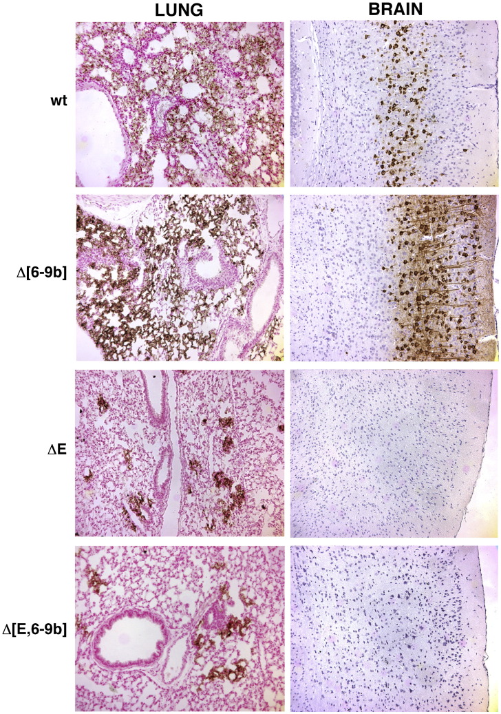
Recombinant severe acute respiratory virus (SARS-CoV) variants lacking the group specific genes 6, 7a, 7b, 8a, 8b and 9b (rSARS-CoV-Delta[6-9b]), the structural gene E (rSARS-CoV-DeltaE), and a combination of both sets of genes (rSARS-CoV-Delta[E,6-9b]) have been generated. All these viruses were rescued in monkey (Vero E6) cells and were also infectious for human (Huh-7, Huh7.5.1 and CaCo-2) cell lines and for transgenic (Tg) mice expressing the SARS-CoV receptor human angiotensin converting enzyme-2 (hACE-2), indicating that none of these proteins is essential for the viral cycle. Furthermore, in Vero E6 cells, all the viruses showed the formation of particles with the same morphology as the wt virus, indicating that these proteins do not have a high impact in the final morphology of the virions. Nevertheless, in the absence of E protein, release of virus particles efficacy was reduced. Viruses lacking E protein grew about 100-fold lower than the wt virus in lungs of Tg infected mice but did not grow in the brains of the same animals, in contrast to the rSARS-CoV-Delta[6-9b] virus, which grew almost as well as the wt in both tissues. Viruses lacking E protein were highly attenuated in the highly sensitive hACE-2 Tg mice, in contrast to the minimal rSARS-CoV-Delta[6-9b] and wt viruses. These data indicate that E gene might be a virulence factor influencing replication level, tissue tropism and pathogenicity of SARS-CoV, suggesting that DeltaE attenuated viruses are promising vaccine candidates.


Figures

Fig. 1
Fig. 1
Rescue of SARS-CoV deletion mutants. Recombinant viruses were rescued by transfecting SARS-CoV cDNA into Vero E6 cells. (A) Genetic organization of the rSARS-CoV-Δ[6–9b] virus. The 912-nt deletion that includes ORFs 6, 7a, 7b, 8a, and the 5′ end of ORF 8b is indicated in a light grey box. ORF 9b (darker grey box) was mutated by changes in the ATG codon and in two in phase downstream ATGs and by the introduction of a stop codon (changes are shown in grey and italics). Letters and numbers indicate the viral genes. L, leader sequence; An, polyA tail. The mutations introduced to delete gene E were previously described (DeDiego et al., 2007). (B) Plaques produced by the indicated viruses on Vero E6 cells.
Fig. 2
Fig. 2
Growth kinetics of the defective viruses in monkey and human cells. Vero E6 (A), CaCo-2 (B), and Huh7.5.1 cells (C) were infected at a moi of 0.05 with the indicated viruses, and viral titers in cell supernatants at different times post-infection were measured by plaque assay on Vero E6 cells. Error bars represent standard deviations of the mean from three experiments.
Fig. 3
Fig. 3
Ultrastructural analysis of infected Vero E6 cells. Vero E6 cells were infected at a moi of 0.5 with SARS-CoV (A), rSARS-CoV-Δ[6–9b] (B), rSARS-CoV-ΔE (C), and rSARS-CoV-Δ[E,6–9b] (D). At 24 h post-infection the cells were processed for electron microscopy of ultrathin sections. Mature virus particles (arrows) were detected in ERGIC sites and swollen Golgi sacs that appeared as large vacuoles. Bars, 200 μm.
Fig. 4
Fig. 4
Morphology of extracellular SARS-CoV deletion mutants. (A) Electron micrographs of ultrathin sections showing extracellular viruses adjacent to the surface of cells infected with the indicated virus. Bars, 200 nm. (B) Ultrathin sections showing the budding process for viruses with (left) and without (right) E protein. Bars, 50 nm.
Fig. 5
Fig. 5
Virulence of the defective viruses. hACE2 Tg mice were intranasally infected with 12,000 pfu (A and B) or with the indicated doses (C and D) of rSARS-CoV, rSARS-CoV-Δ[6–9b], rSARS-CoV-ΔE and rSARS-CoV-Δ[E,6–9b] viruses. Animals were monitored daily for weight (A) and mortality (B). To further analyze the virulence of rSARS-CoV-Δ[6–9b], hACE2 Tg mice were infected with the indicated doses of rSARS-CoV or rSARS-CoV-Δ[6–9b] and weight (C) and mortality (D) monitored.
Fig. 6
Fig. 6
In vivo growth kinetics of the variant viruses. hACE2 Tg mice were intranasally inoculated with 12,000 pfu. At 2 and 4 days post-infection, lung (A) and brain (B) tissues were harvested and viral titers were analyzed in Vero E6 cell monolayers. Numbers over bars indicate numbers of mice with detectable virus in relation to the total number of examined mice. Absence of numbers indicate that virus was detected in all examined animals. Black bars, SARS-CoV; gray bars, rSARS-CoV-Δ[6–9b], black dashed bars, rSARS-CoV-ΔE, and gray dashed bars, rSARS-CoV-Δ[E, 6–9b].
Fig. 7
Fig. 7
Histopathology and immunohistochemistry. hACE2 Tg mice were intranasally inoculated with 12,000 pfu of the indicated viruses and sacrificed at day 4 p.i. Zinc formalin-fixed lungs (left) and brain (cerebrum) (right) were analyzed for viral antigen as described in Materials and methods. Original magnification was 10×.

Similar articles

Cited by

References

    1. Almazan F., Galan C., Enjuanes L. The nucleoprotein is required for efficient coronavirus genome replication. J. Virol. 2004;78:12683–12688. - PMC - PubMed
    1. Almazan F., DeDiego M.L., Galan C., Escors D., Alvarez E., Ortego J., Sola I., Zuñiga S., Alonso S., Moreno J.L., Nogales A., Capiscol C., Enjuanes L. Construction of a SARS-CoV infectious cDNA clone and a replicon to study coronavirus RNA synthesis. J. Virol. 2006;80:10900–10906. - PMC - PubMed
    1. Chen C.Y., Ping Y.H., Lee H.C., Chen K.H., Lee Y.M., Chan Y.J., Lien T.C., Jap T.S., Lin C.H., Kao L.S., Chen Y.M. Open reading frame 8a of the human severe acute respiratory syndrome coronavirus not only promotes viral replication but also induces apoptosis. J. Infect. Dis. 2007;196:405–415. - PMC - PubMed
    1. Corse E., Machamer C.E. Infectious bronchitis virus E protein is targeted to the Golgi complex and directs release of virus-like particles. J. Virol. 2000;74:4319–4326. - PMC - PubMed
    1. Corse E., Machamer C.E. The cytoplasmic tail of infectious bronchitis virus E protein directs Golgi targeting. J. Virol. 2002;76:1273–1284. - PMC - PubMed

Publication types