

Staphylococcal biofilms
- PMID: 18453278
- PMCID: PMC2777538
- DOI: 10.1007/978-3-540-75418-3_10
Staphylococcal biofilms
Abstract
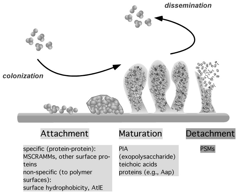
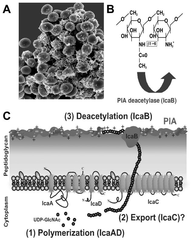
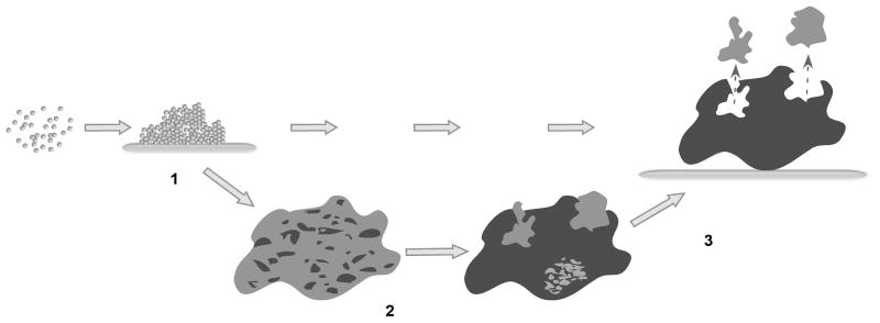
Staphylococcus epidermidis and Staphylococcus aureus are the most frequent causes of nosocomial infections and infections on indwelling medical devices, which characteristically involve biofilms. Recent advances in staphylococcal molecular biology have provided more detailed insight into the basis of biofilm formation in these opportunistic pathogens. A series of surface proteins mediate initial attachment to host matrix proteins, which is followed by the expression of a cationic glucosamine-based exopolysaccharide that aggregates the bacterial cells. In some cases, proteins may function as alternative aggregating substances. Furthermore, surfactant peptides have now been recognized as key factors involved in generating the three-dimensional structure of a staphylococcal biofilm by cell-cell disruptive forces, which eventually may lead to the detachment of entire cell clusters. Transcriptional profiling experiments have defined the specific physiology of staphylococcal biofilms and demonstrated that biofilm resistance to antimicrobials is due to gene-regulated processes. Finally, novel animal models of staphylococcal biofilm-associated infection have given us important information on which factors define biofilm formation in vivo. These recent advances constitute an important basis for the development of anti-staphylococcal drugs and vaccines.
Figures
References
-
- Arber N, et al. Pacemaker endocarditis. Report of 44 cases and review of the literature. Medicine (Baltimore) 1994;73:299–305. - PubMed
-
- Arciola CR, An YH, Campoccia D, Donati ME, Montanaro L. Etiology of implant orthopedic infections: a survey on 1027 clinical isolates. Int J Artif Organs. 2005;28:1091–1100. - PubMed
-
- Arciola CR, et al. Detection of biofilm formation in Staphylococcus epidermidis from implant infections. Comparison of a PCR-method that recognizes the presence of ica genes with two classic phenotypic methods. J Biomed Mater Res A. 2006;76:425–430. - PubMed
-
- Balaban N, et al. Use of the quorum-sensing inhibitor RNAIII-inhibiting peptide to prevent biofilm formation in vivo by drug-resistant Staphylococcus epidermidis. J Infect Dis. 2003;187:625–630. - PubMed
Publication types
MeSH terms
Grants and funding
LinkOut - more resources
Full Text Sources
Other Literature Sources
Medical
