

Yersinia pestis biofilm in the flea vector and its role in the transmission of plague
- PMID: 18453279
- PMCID: PMC3727414
- DOI: 10.1007/978-3-540-75418-3_11
Yersinia pestis biofilm in the flea vector and its role in the transmission of plague
Abstract
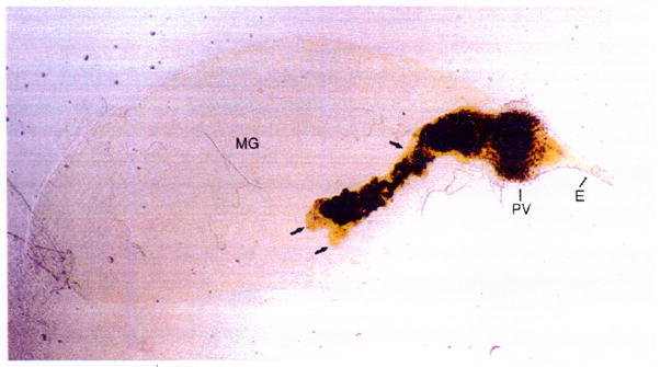
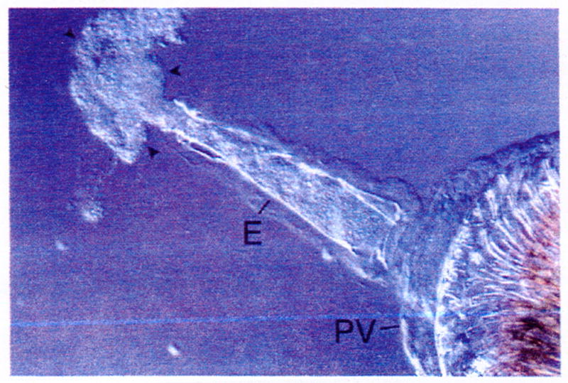
Transmission by fleabite is a relatively recent evolutionary adaptation of Yersinia pestis, the bacterial agent of bubonic plague. To produce a transmissible infection, Y. pestis grows as an attached biofilm in the foregut of the flea vector. Biofilm formation both in the flea foregut and in vitro is dependent on an extracellular matrix (ECM) synthesized by the Yersinia hms gene products. The hms genes are similar to the pga and ica genes of Escherichia coli and Staphylococcus epidermidis, respectively, that act to synthesize a poly-beta-1,6-N-acetyl-d-glucosamine ECM required for biofilm formation. As with extracellular polysaccharide production in many other bacteria, synthesis of the Hms-dependent ECM is controlled by intracellular levels of cyclic-di-GMP. Yersinia pseudotuberculosis, the food- and water-borne enteric pathogen from which Y. pestis evolved recently, possesses identical hms genes and can form biofilm in vitro but not in the flea. The genetic changes in Y. pestis that resulted in adapting biofilm-forming capability to the flea gut environment, a critical step in the evolution of vector-borne transmission, have yet to be identified. During a flea bite, Y. pestis is regurgitated into the dermis in a unique biofilm phenotype, and this has implications for the initial interaction with the mammalian innate immune response.
Figures
References
-
- Achtman M, Morelli G, Zhu P, Wirth T, Diehl I, Kusecek B, Vogler AJ, Wagner DM, Allender CJ, Easterday WR, Chenal-Francisque V, Worsham P, Thomson NR, Parkhill J, Lindler LE, Carniel E, Keim P. Microevolution and history of the plague bacillus, Yersinia pestis. Proc Natl Acad Sci U S A. 2004;101:17837–17842. - PMC - PubMed
Publication types
MeSH terms
Grants and funding
LinkOut - more resources
Full Text Sources
Other Literature Sources
Medical
Research Materials
Miscellaneous
