. 2009 Jan;19(1):176-86.
doi: 10.1093/cercor/bhn068. Epub 2008 May 2.

Visuokinesthetic perception of hand movement is mediated by cerebro-cerebellar interaction between the left cerebellum and right parietal cortex

Affiliations

Visuokinesthetic perception of hand movement is mediated by cerebro-cerebellar interaction between the left cerebellum and right parietal cortex

Nobuhiro Hagura et al. Cereb Cortex. 2009 Jan.

Abstract

Combination of visual and kinesthetic information is essential to perceive bodily movements. We conducted behavioral and functional magnetic resonance imaging experiments to investigate the neuronal correlates of visuokinesthetic combination in perception of hand movement. Participants experienced illusory flexion movement of their hand elicited by tendon vibration while they viewed video-recorded flexion (congruent: CONG) or extension (incongruent: INCONG) motions of their hand. The amount of illusory experience was graded by the visual velocities only when visual information regarding hand motion was concordant with kinesthetic information (CONG). The left posterolateral cerebellum was specifically recruited under the CONG, and this left cerebellar activation was consistent for both left and right hands. The left cerebellar activity reflected the participants' intensity of illusory hand movement under the CONG, and we further showed that coupling of activity between the left cerebellum and the "right" parietal cortex emerges during this visuokinesthetic combination/perception. The "left" cerebellum, working with the anatomically connected high-order bodily region of the "right" parietal cortex, participates in online combination of exteroceptive (vision) and interoceptive (kinesthesia) information to perceive hand movement. The cerebro-cerebellar interaction may underlie updating of one's "body image," when perceiving bodily movement from visual and kinesthetic information.


Figures
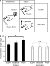
Figure 1.
Figure 1.
Conditions (a) and results (b) in the behavioral experiment. (a) Participants experienced illusory flexions of their right hands while viewing their video-recorded hand flexion (CONG) or extension motion (INCONG). Crosses on the wrist joints indicate fixation points. The open arrow indicates the direction of illusory movement, and filled arrows indicate the directions of visual hand motions. Three different velocities were used for each hand motion. (b) Filled bars represent the mean illusory angles across all participants under CONG conditions, and open bars indicate those under INCONG conditions. Error bars indicate standard errors of means across all participants. *P < 0.05.
Figure 2.
Figure 2.
Results from fMRI experiment 1 and 2. (ad) Activations in the motor areas and right-dominant frontoparietal areas during visuokinesthetic processing under the CONG and INCONG conditions. (e) Left cerebellar activations exclusively under the CONG condition. The orange region indicates the active area in fMRI experiment 1 (right hand), and the blue region corresponds to that identified in fMRI experiment 2 (activated for the left and right hands), respectively. The horizontal plane (z = −27) is displayed. (f) The size of effects of left cerebellar activation across conditions in the first fMRI experiment. Bars indicate the means of contrast parameter estimates (size of effect in arbitrary units) for left cerebellar activation (−27, −69, −30) during CONG (orange bar), INCONG (black bar), and control conditions (see Materials and Methods). The size of effect was calculated by comparing each condition with the condition in which participants simply viewed the fixation point (Fi–N; see Materials and Methods). Error bars represent standard errors of means across participants.
Figure 3.
Figure 3.
Results from fMRI experiment 3. (a) Mean illusion scores in fMRI experiment 3. **P < 0.01, ***P < 0.001. Error bars indicate the standard errors of means across participants. (b) The left cerebellar region (red region; P < 0.005, uncorrected for display purpose) in which activity was correlated with the intensity of visuokinesthetic perception. The activation is superimposed on the same plane as that in Figure 2e. (c) Significant correlation between behavioral ratings (illusion scores) and left cerebellar activity (size of effect) in a representative participant (r = 0.57, df = 34, P < 0.001, 1-tailed). The illusion scores are normalized (mean corrected) in each session.
Figure 4.
Figure 4.
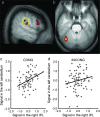
Results from flip analysis (a) and from PPI analyses (bd). (a) Right-dominant activities in the IPL (yellow circle) and in areas 44/45 revealed by the main effect of illusions. A sagittal plane (x = 60) is displayed. (b) Left cerebellar activation of a representative participant, which showed enhanced coupling of activity with right IPL activity under the CONG condition (P < 0.005, uncorrected for display purpose). A horizontal plane (z = −27) is displayed. (c) and (d) Relationship of activities between the right IPL and the left cerebellum in the representative participant (c: CONG; d: INCONG). The regression slopes were 0.52 and 0.29 for the CONG and INCONG conditions, respectively. The activities (x-axis for right IPL; y-axis for left cerebellum) are mean adjusted (arbitrary units).

References

    1. Allen G, Buxton RB, Wong EC, Courchesne E. Attentional activation of the cerebellum independent of motor involvement. Science. 1997;275:1940–1943. - PubMed
    1. Allin M, Nosarti C, Rifkin L, Wyatt J, Murray R. Visuomotor integration and the cerebellum in adolescents born preterm. Neuroimage. 2005;26:S23. - PubMed
    1. Bard C, Fleury M, Teasdale N, Paillard J, Nougier V. Contribution of proprioception for calibrating and updating the motor space. Can J Physiol Pharmacol. 1995;73:246–254. - PubMed
    1. Bauswein E, Kolb FP, Leimbeck B, Rubia FJ. Simple and complex spike activity of cerebellar Purkinje cells during active and passive movements in the awake monkey. J Physiol. 1983;339:379–394. - PMC - PubMed
    1. Beppu H, Suda M, Tanaka R. Analysis of cerebellar motor disorders by visually guided elbow tracking movement. Brain. 1984;107:787–809. - PubMed

Publication types