. 2008 Sep;29(9):1717-24.
doi: 10.1093/carcin/bgn098. Epub 2008 May 2.

Abi1 gene silencing by short hairpin RNA impairs Bcr-Abl-induced cell adhesion and migration in vitro and leukemogenesis in vivo

Affiliations

Abi1 gene silencing by short hairpin RNA impairs Bcr-Abl-induced cell adhesion and migration in vitro and leukemogenesis in vivo

Weidong Yu et al. Carcinogenesis. 2008 Sep.

Abstract

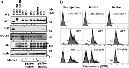
Abl interactor (Abi) 1 was first identified as the downstream target of Abl tyrosine kinases and was found to be dysregulated in leukemic cells expressing oncogenic Bcr-Abl and v-Abl. Although the accumulating evidence supports a role of Abi1 in actin cytoskeleton remodeling and growth factor/receptor signaling, it is not clear how it contributes to Bcr-Abl-induced leukemogenesis. We show here that Abi1 gene silencing by short hairpin RNA attenuated the Bcr-Abl-induced abnormal actin remodeling, membrane-type 1 metalloproteinase clustering and inhibited cell adhesion and migration on fibronectin-coated surfaces. Although the knock down of Abi1 expression did not affect growth factor-independent growth of Bcr-Abl-transformed Ba/F3 cells in vitro, it impeded competitive expansion of these cells in non obese diabetic (NOD)/ severe combined immuno-deficiency (SCID) mice. Remarkably, the knock down of Abi1 expression in Bcr-Abl-transformed Ba/F3 cells impaired the leukemogenic potential of these cells in NOD/SCID mice. Abi1 contributes to Bcr-Abl-induced leukemogenesis in part through Src family kinases, as the knock down of Abi1 expression attenuates Bcr-Abl-stimulated activation of Lyn. Together, these data provide for the first time the direct evidence that supports a critical role of Abi1 pathway in the pathogenesis of Bcr-Abl-induced leukemia.


Figures

Fig. 1.
Fig. 1.
The shRNA-mediated Abi1 gene silencing. (A) Abi1 expression in Ba/F3 cells (BaF3) and p185wt cells transduced with either non-silencing shRNA (p185wt) or Abi1 shRNAs (shRNAs A, B and C). Total lysates from 1 × 10⁶ cells were separated on sodium dodecyl sulfate–polyacrylamide gel electrophoresis and were transferred to nitrocellulose membrane. The membrane was probed with the antibodies as indicated. The p185 Bcr-Abl and c-Abl were indicated by the arrow and arrowhead, respectively. (B) Efficacy of Abi1 knockdown by different Abi1 shRNAs. The lysates (2 × 10⁷ cells) from the indicated cell lines were immunoprecipitated (IP) by Abi1 antibody and the immunoprecipitates were analyzed by western blotting (WB) using Abi1 antibody as probe (upper panel). Total RNAs from the indicated cell lines were isolated and the complementary DNAs were synthesized. Real-time quantitative PCR (lower panel) was performed to determine Abi1 messenger RNA (mRNA) levels, which are expressed as the levels relative to that of GAPDH. Data represent mean ± SD of triplicate experiments; *P < 0.001. (C) Expression of Abi2 and WAVE2 in Ba/F3 cells and p185wt cells transduced with either non-silencing shRNA or Abi1 shRNAs. Total lysates from 1 × 10⁶ cells were separated on sodium dodecyl sulfate–polyacrylamide gel electrophoresis and were transferred to nitrocellulose membrane. The membrane was probed with the antibodies as indicated.
Fig. 2.
Fig. 2.
Abi1 knockdown did not affect Bcr-Abl-induced protein tyrosine phosphorylation and IL3-independent growth. (A) Profiles of protein tyrosine phosphorylation in Ba/F3 cells and p185wt cells transduced with either non-silencing shRNA (p185wt) or Abi1 shRNAs (Abi1R). Indicated cell lines were starved in RPMI 1640 plus 0.1% bovine serum albumin for 6 h prior to harvest. Total lysates from 1 × 10⁶ cells were separated on 8% sodium dodecyl sulfate–polyacrylamide gel electrophoresis and subjected to western blot analysis with anti-phosphotyrosine antibody. (B) IL3-independent growth of Ba/F3 cells as well as the p185wt cells transduced with either non-silencing shRNA (p185wt) or Abi1 shRNA (Abi1R).
Fig. 3.
Fig. 3.
Abi1 knockdown inhibited Bcr-Abl-stimulated actin cytoskeleton remodeling, MT1-MMP clustering, as well as cell adhesion and migration on fibronectin-coated surfaces. (A) Inhibition of Bcr-Abl-induced abnormal actin remodeling by Abi1 knockdown. Ba/F3 cells and the Ba/F3 cells expressing p185wt, p185K671R and p185wt plus Abi1 shRNA were fixed and stained with TRITC-conjugated phalloidin. The cells with F-actin-rich structures (invadopodia-like structures) were visualized by fluorescence microscopy as shown by arrowheads (upper panel) and were counted (lower panel, represented as mean ± SD percentage of three randomly picked areas). (B) The p185wt cells expressing GFP–MT1-MMP were transduced with either control retrovirus (control) or the retrovirus expressing Abi1 shRNA. The knock down of Abi1 expression was confirmed by western blotting (data not shown). The distribution of GFP–MT1-MMP was visualized by fluorescence microscopy. A similar result has been described previously (41). (C) Effects of Abi1 knockdown on Bcr-Abl-stimulated cell adhesion (lower panel) and migration (upper panel) on fibronectin-coated surfaces. Ba/F3 cells and the p185wt cells transduced with either non-silencing shRNA or Abi1 shRNAs were grown in fibronectin-coated six-well plate (2.5 × 10⁵ per well) for 16 h. The total cells and the cells that were adherent to fibronectin-coated surfaces were counted and the percentage of adherent cells calculated. The vertical axis shows the percentage of the adherent cells and is expressed as the mean ± SD of triplicate wells. For cell migration on fibronectin-coated membrane, 1 × 10⁵ cells were tested in Transwell migration assay. The vertical axis shows the percentage of the migrated cells and is expressed as the mean ± SD of triplicate wells; *P < 0.01.
Fig. 4.
Fig. 4.
Bcr-Abl-induced leukemogenesis was impaired by Abi1 knockdown. (A) Stable knock down of Abi1 expression in p185wt cells transduced with Abi1 shRNA. The Ba/F3 cells and the p185wt cells transduced with a non-silencing shRNA or Abi1 shRNA were grown in vitro for indicated time. Total lysates from 1 × 10⁶ cells were separated on sodium dodecyl sulfate–polyacrylamide gel electrophoresis, transferred to nitrocellulose membrane and subjected to western blot analysis using indicated antibodies. (B) Survival of the mice injected with Ba/F3 cells, control p185wt cells and the p185wt cells expressing Abi1 shRNA.
Fig. 5.
Fig. 5.
Pathology analysis of the mice injected with Ba/F3 cells, control p185wt cells and p185wt cells transduced with Abi1 shRNA. (A) Spleen weight of mice injected with Ba/F3 cells (control) and the p185wt cells expressing with (p185wt + Abi1 shRNA) or without (p185wt) Abi1 shRNA. (B) Histology of spleens and livers from the mice receiving Ba/F3 cells (control) and the p185wt cells expressing with (p185wt + Abi1 shRNA) or without (p185wt ctrl) Abi1 shRNA. Arrowheads indicate the massively expanded p185wt cells.
Fig. 6.
Fig. 6.
Abi1 knockdown impeded in vivo competitive expansion of p185wt cells. (A) Expression of Abi1 in Ba/F3 cells, p185wt cells transduced with non-silencing shRNA (p185wt control) or Abi1 shRNA (p185wt Abi1 Ri), as well as the cells rescued from bone marrow (BM), peripheral blood (PB) and spleen of the diseased mice injected with p185wt cells transduced with either non-silencing shRNA or Abi1 shRNA. The 100 μg total proteins from these cells were separated by sodium dodecyl sulfate–polyacrylamide gel electrophoresis, transferred to nitrocellulose membrane and western blotted (WB) with indicated antibodies. Molecular markers are indicated. The arrow indicates p185Bcr-Abl. (B) In vivo competitive expansion of p185wt GFP cells and p185wt Abi1 shRNA cells. The p185wt cells were transduced with MSCV-based retroviruses expressing either GFP or Abi1 shRNA. The GFP-positive p185wt cells were sorted by fluorescence-activated cell sorting to a purity of >95%. The GFP-positive cells then were mixed with p185wt cells expressing Abi1 shRNA at a ratio of 1:1. The mixed cells were either cultured in vitro for 5 weeks or injected into Balb/c mice (1 × 10⁶ cells per mouse) through tail vein. The cells derived from p185wt cells were rescued from bone marrow and peripheral blood (data not shown) of the diseased mice by selection with puromycin. The rescued cells and the cells cultured in vitro were subjected to flow cytometry analysis.
Fig. 7.
Fig. 7.
The knock down of Abi1 expression attenuated Bcr-Abl-induced activation of Lyn kinases. Ba/F3 cells (lanes 1 and 4), control p185wt cells (lanes 2 and 5) and p185wt Abi1 shRNA cells (lanes 3 and 6) were starved in RPMI 1640 plus 0.1% bovine serum albumin for 6 h. The cells were then treated without (lanes 1–3) or with (lanes 4–6) recombinant rat IL3 (20 ng/ml) for 10 min. The lysates (2 × 10⁷ cells) from indicated cell lines were immunoprecipitated (IP) with anti-Lyn antibodies, separated on 8% sodium dodecyl sulfate–polyacrylamide gel electrophoresis and subjected to western blot (WB) analysis using anti-Src p416 antibody, which recognizes activated Lyn (upper panel). The membrane then was stripped and reprobed with anti-Lyn antibodies (lower panel). Two Lyn isoforms, p53Lyn and p56Lyn, were indicated.

References

    1. Druker BJ, et al. Chronic myelogenous leukemia. Hematology Am Soc Hematol Educ Program. 2001:87–112. - PubMed
    1. Melo JV, et al. Chronic myeloid leukaemia as a model of disease evolution in human cancer. Nat. Rev. Cancer. 2007;7:441–453. - PubMed
    1. Hehlmann R, et al. Chronic myeloid leukaemia. Lancet. 2007;370:342–350. - PubMed
    1. Ren R. Mechanisms of BCR-ABL in the pathogenesis of chronic myelogenous leukaemia. Nat. Rev. Cancer. 2005;5:172–183. - PubMed
    1. Van Etten RA. Oncogenic signaling: new insights and controversies from chronic myeloid leukemia. J. Exp. Med. 2007;204:461–465. - PMC - PubMed

Publication types

MeSH terms

LinkOut - more resources