. 2008 May 15;180(10):6457-61.
doi: 10.4049/jimmunol.180.10.6457.

Cutting edge: central nervous system plasmacytoid dendritic cells regulate the severity of relapsing experimental autoimmune encephalomyelitis

Affiliations

Cutting edge: central nervous system plasmacytoid dendritic cells regulate the severity of relapsing experimental autoimmune encephalomyelitis

Samantha L Bailey-Bucktrout et al. J Immunol. .

Abstract

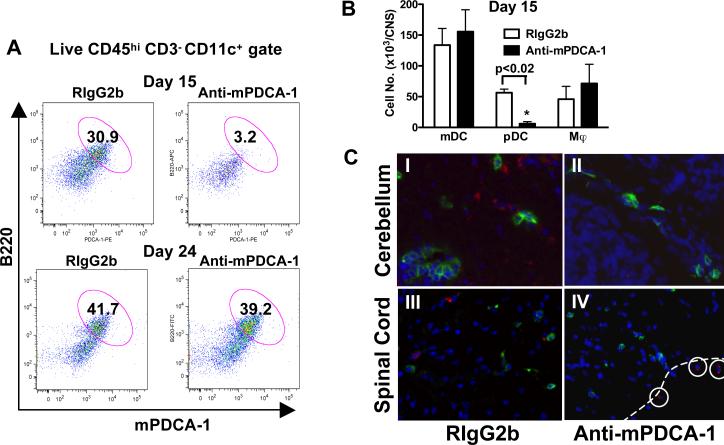
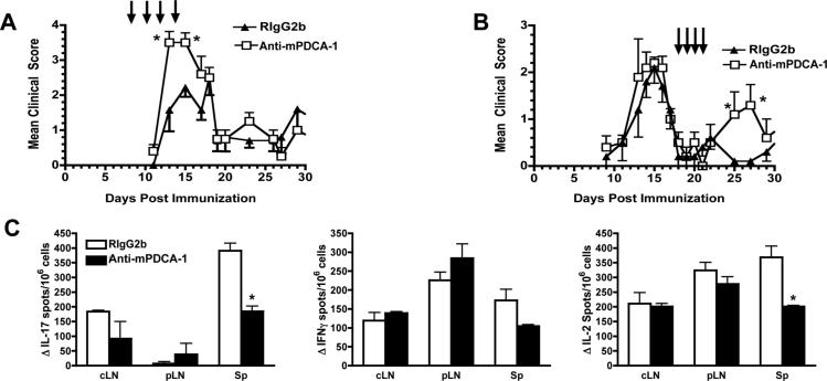
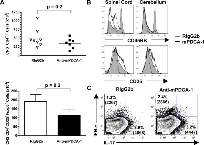
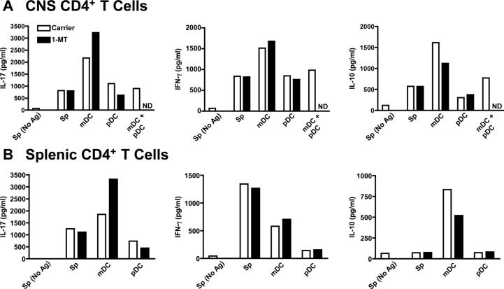
Plasmacytoid dendritic cells (pDCs) have both stimulatory and regulatory effects on T cells. pDCs are a major CNS-infiltrating dendritic cell population during experimental autoimmune encephalomyelitis but, unlike myeloid dendritic cells, have a minor role in T cell activation and epitope spreading. We show that depletion of pDCs during either the acute or relapse phases of experimental autoimmune encephalomyelitis resulted in exacerbation of disease severity. pDC depletion significantly enhanced CNS but not peripheral CD4(+) T cell activation, as well as IL-17 and IFN-gamma production. Moreover, CNS pDCs suppressed CNS myeloid dendritic cell-driven production of IL-17, IFN-gamma, and IL-10 in an IDO-independent manner. The data demonstrate that pDCs play a critical regulatory role in negatively regulating pathogenic CNS CD4(+) T cell responses, highlighting a new role for pDCs in inflammatory autoimmune disease.


Figures

Figure 1
Figure 1. anti-mPDCA-1 efficiently and specifically depletes CNS pDCs during EAE
(a) SJL mice were treated with anti-mPDCA-1 or isotype-matched control (Rat IgG2b) mAb on days 8-14 after EAE induction. One day (day 15) and 9 days after pDC depletion (day 24), the effect of treatment on gated live CNS DCs (CD45highCD3CD11c+) was determined. (b) Day 15 analysis of CNS CD11b+ mDCs, CD11bB220+ pDCs, and CD11b+CD11c macrophages (Mϕ). Data are mean ± SEM of 4 animals per group representative of 5 experiments. p value determined by Mann-Whitney. (c) Day 15 immunohistochemical analysis of (a), cerebellum (I and II) and lumbar spinal cord (III and IV) stained for mPDCA-1 (red), CD4 (green) and DAPI (blue). Dotted line shows the meningeal edge of spinal cord tissue. mPDCA-1+ pDCs are circled. Magnifications ×200 (I and II) and ×100 (III and IV) are representative of 3 mice.
Figure 2
Figure 2. CNS pDC depletion increases the severity of acute and relapsing R-EAE
(a) Clinical EAE course following anti-mPDCA-1 or isotype control (RIgG2b) mAb treatment (arrows). Mean clinical scores ± SEM of 5-7 mice per treatment group, 1 of 5 experiments. (b) Treatment of EAE during remission (days 18-21). Mean clinical scores + SEM of 5 mice per treatment group, 1 of 2 experiments. *Mean clinical scores are significantly different from controls (p < 0.05; Mann-Whitney). c) Day 15 ELISPOTS for IFN-γ, IL-17, and IL-2 in response to PLP139-151 with cells from the cervical LN (cLN), inguinal LN (pLN) and spleen (Sp). Mean ± SEM of 3 animals, 1 of 3experiments. *Values significantly different, p < 0.01; un-paired Students t test.
Figure 3
Figure 3. pDCs regulate CNS CD4+ T cell activation and IFN-γ and IL-17 production
(a) At day 15 after EAE induction, and treatment with RIgG2b or mPDCA-1 days 8-14, the numbers of CNS CD45+CD3+CD4+ T cells and CD4+FoxP3+CD25+ Tregs were enumerated. Data points for CD4+ T cells represents mice from 2 experiments, and the bars indicate the number of FoxP3+ T cells ± SEM of 4 animals in 2 experiments. p values determined by Mann-Whitney. (b) CD45RB and CD25 expression by CNS CD4+ T cells at day 15. Isotype control staining is shown in dotted lines of corresponding colors. Representative of 4 animals from 2 experiments. (c) Intracellular cytokine analysis of CNS CD4+ T cells isolated from 2 mice at day 15. The percentage of IFN-γ and IL-17 staining cells within the viable CD45hiCD3+CD4+ gate and the mean fluorescence intensities are indicated in parentheses. Results representative of 3 experiments.
Figure 4
Figure 4. pDCs actively suppress mDC supported IFN-γ, IL-17 and IL-10 production by CNS CD4+ T cells
On day 14, 105 CD4+ T cells from the CNS (a) or spleen (b) were cultured with 2x10⁴ irradiated splenic APCs (Sp), or sorted CNS pDCs, mDCs or a 1:1 mixture of pDCs and mDCs in the absence (white bars) or presence (black bars) of 1-MT. The amount of secreted IFN-γ, IL-17 and IL-10 in the culture supernatants was determined 4 days later. The results are representative of 3 experiments where 15-20 perfused mice were pooled. ND, not done.

Similar articles

Cited by

References

    1. Hohlfeld R, Wekerle H. Autoimmune concepts of multiple sclerosis as a basis for selective immunotherapy: from pipe dreams to (therapeutic) pipelines. Proc. Natl. Acad. Sci. U. S. A. 2004;101(Suppl 2):14599–14606. - PMC - PubMed
    1. Langrish CL, Chen Y, Blumenschein WM, Mattson J, Basham B, Sedgwick JD, McClanahan T, Kastelein RA, Cua DJ. IL-23 drives a pathogenic T cell population that induces autoimmune inflammation. J. Exp. Med. 2005;201:233–240. - PMC - PubMed
    1. Veldhoen M, Hocking RJ, Flavell RA, Stockinger B. Signals mediated by transforming growth factor-beta initiate autoimmune encephalomyelitis, but chronic inflammation is needed to sustain disease. Nat Immunol. 2006;7:1151–1156. - PubMed
    1. Vanderlugt CL, Miller SD. Epitope spreading in immune-mediated diseases: implications for immunotherapy. Nat. Rev. Immunol. 2002;2:85–95. - PubMed
    1. McMahon EJ, Bailey SL, Castenada CV, Waldner H, Miller SD. Epitope spreading initiates in the CNS in two mouse models of multiple sclerosis. Nat. Med. 2005;11:335–339. - PubMed

Publication types

MeSH terms