. 2008 May 15;180(10):6933-40.
doi: 10.4049/jimmunol.180.10.6933.

Heme oxygenase-1 protects against neutrophil-mediated intestinal damage by down-regulation of neutrophil p47phox and p67phox activity and O2- production in a two-hit model of alcohol intoxication and burn injury

Affiliations

Heme oxygenase-1 protects against neutrophil-mediated intestinal damage by down-regulation of neutrophil p47phox and p67phox activity and O2- production in a two-hit model of alcohol intoxication and burn injury

Xiaoling Li et al. J Immunol. .

Abstract

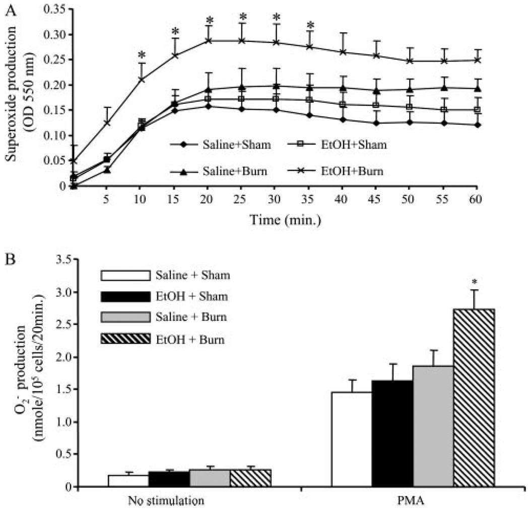
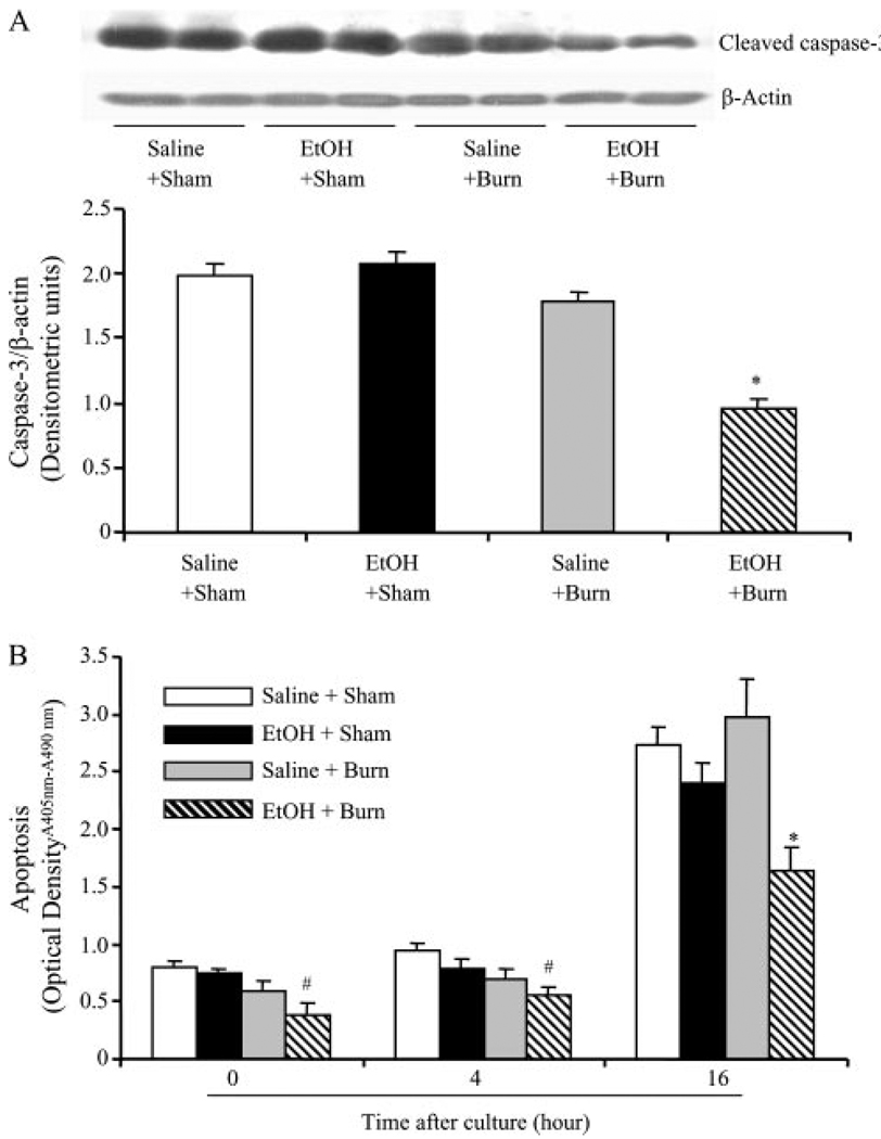
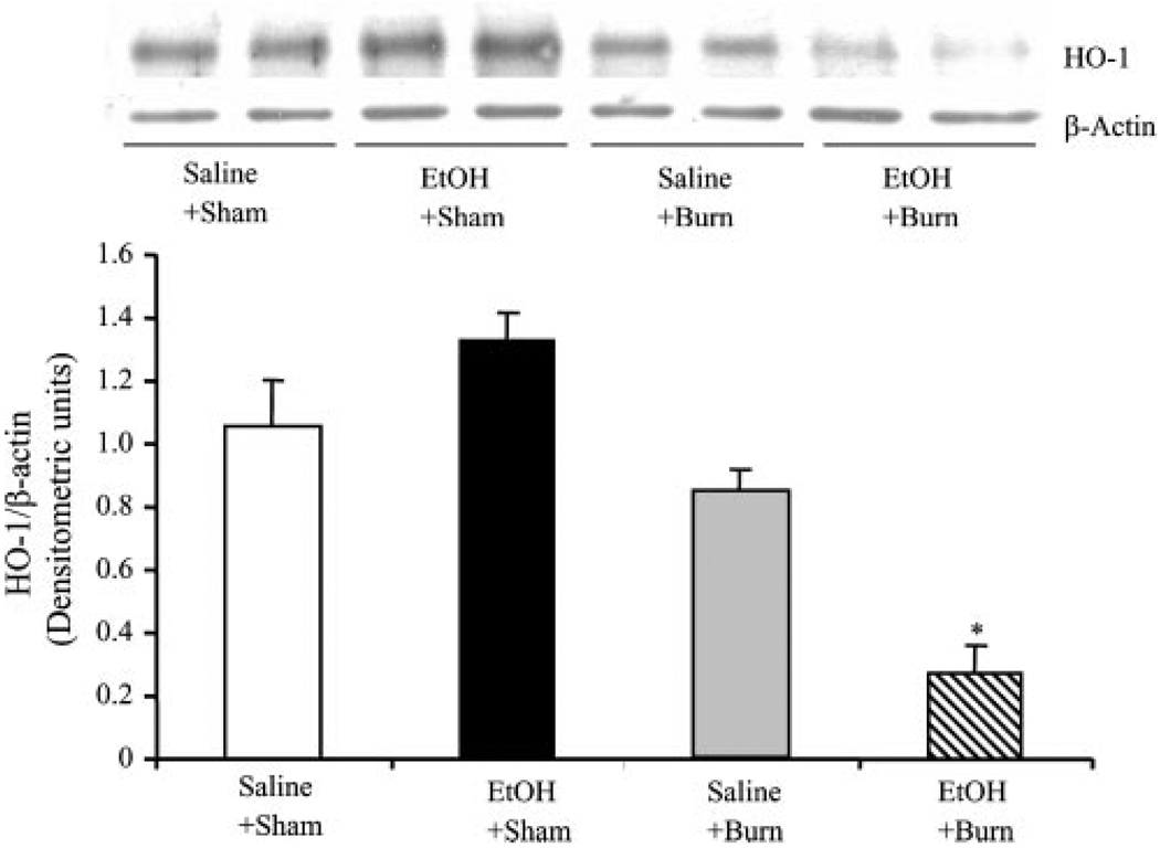
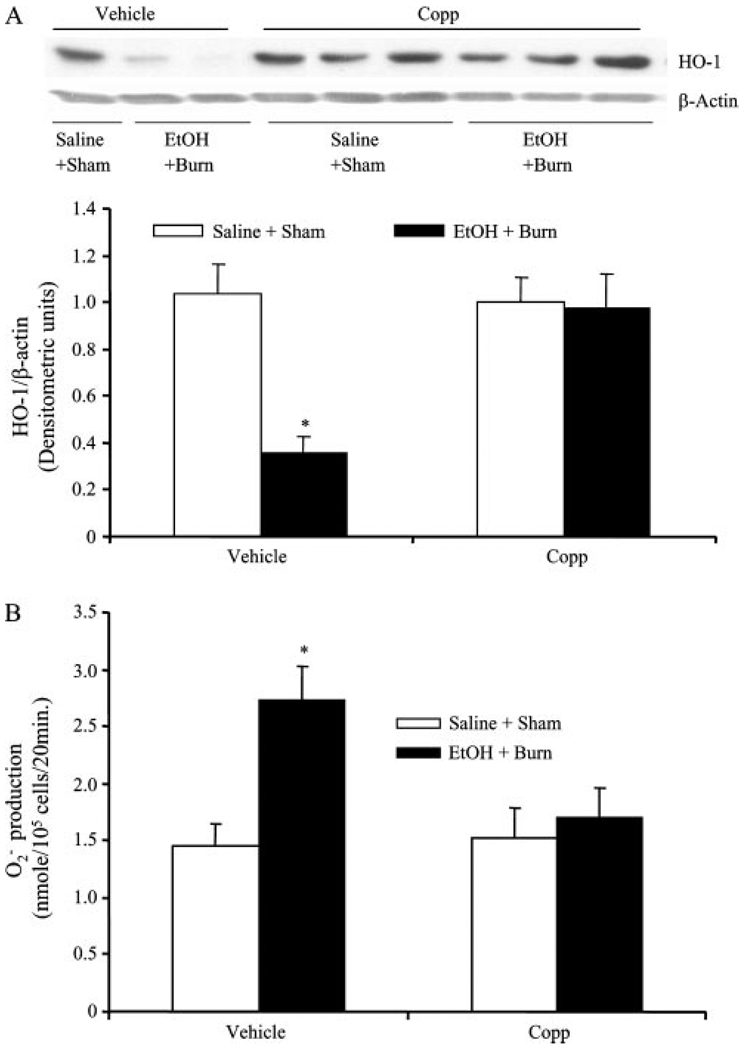
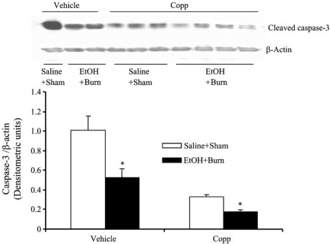
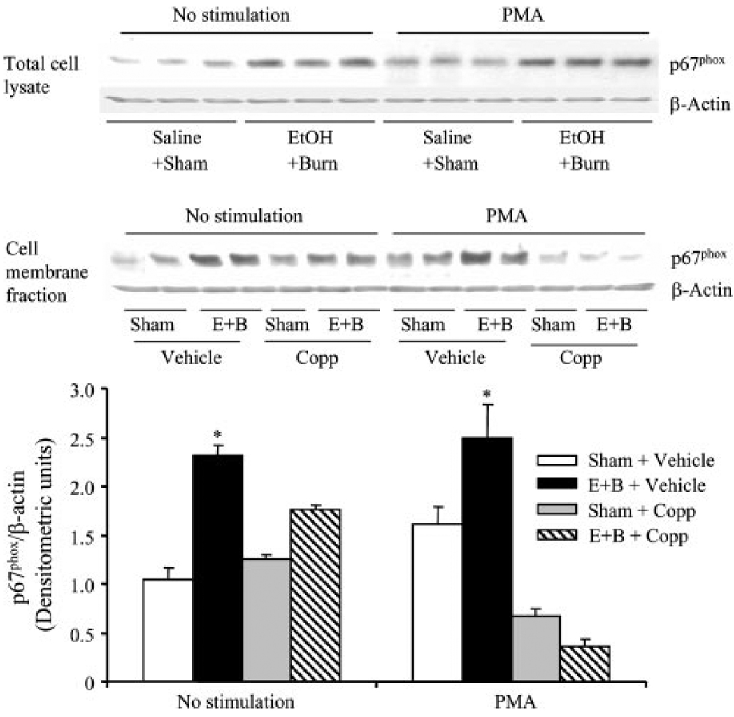
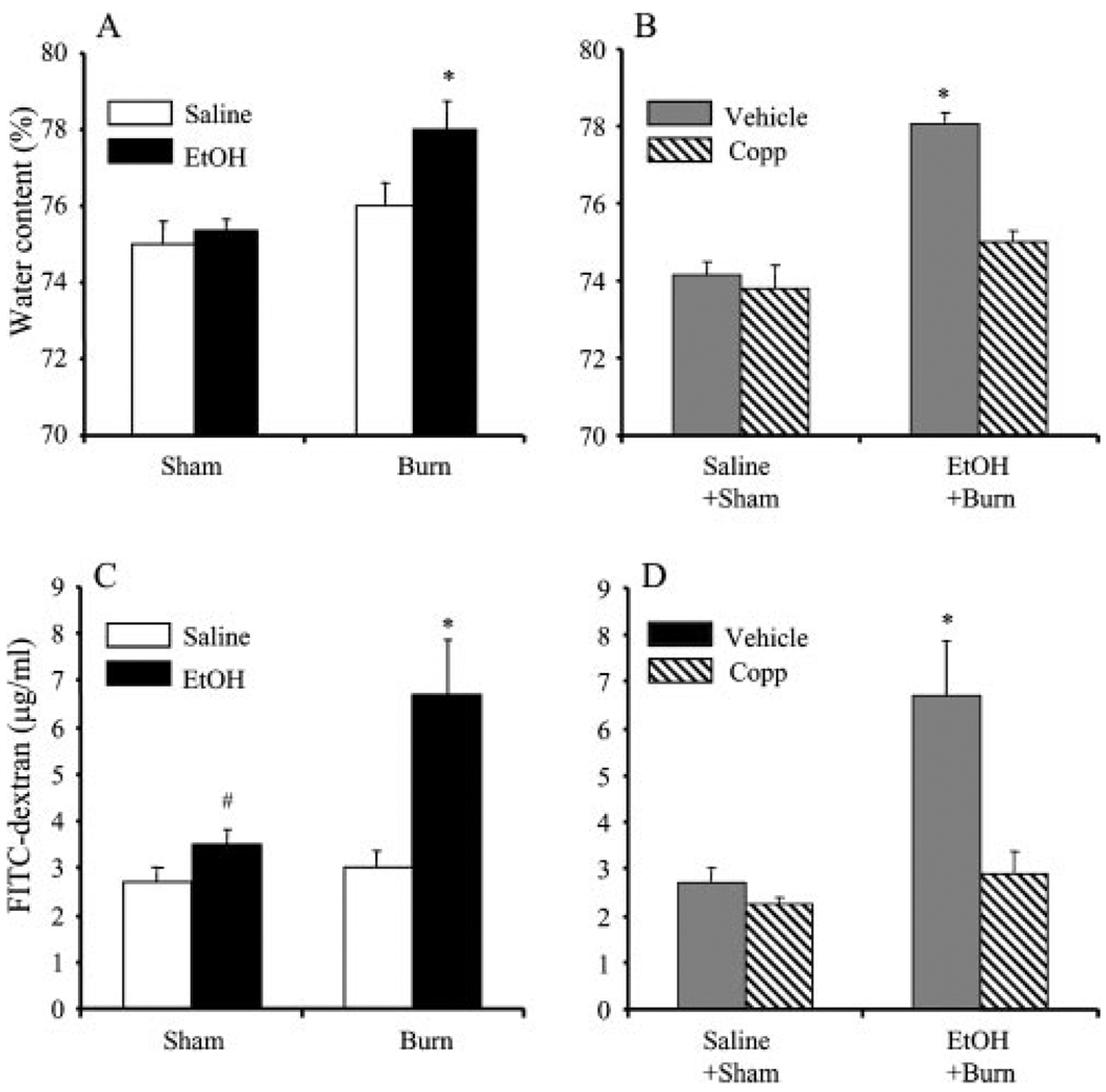
Heme oxygenase-1 (HO-1) has been demonstrated to protect against tissue injury. Furthermore, HO-1 is also shown to be antioxidant. Our recent findings indicate that acute alcohol (EtOH) intoxication exacerbates postburn intestinal and lung tissue damage, and this was found to be neutrophil dependent. Because neutrophil-mediated tissue injury involves the release of superoxide anions (O(2)(-)), the present study examined the role of HO-1 in neutrophil O(2)(-) production following EtOH and burn injury. Furthermore, we investigated whether HO-1 antioxidant properties are mediated via modulation of p47(phox) and/or p67(phox) proteins. Male rats (approximately 250 g) were gavaged with EtOH to achieve a blood EtOH level of approximately 100 mg/dL before burn or sham injury (approximately 12.5% total body surface area). Some rats were treated with HO-1 activator cobalt protoporphyrin IX chloride (Copp; 25 mg/kg body weight) at the time of injury. On day 1 after injury, we found that EtOH combined with burn injury significantly increased neutrophil O(2)(-) production and p47(phox) and p67(phox) activation and decreased caspase-3 activity and apoptosis. This was accompanied with a decrease in neutrophil HO-1 levels. The treatment of animals with HO-1 activator Copp normalized neutrophil HO-1, O(2)(-), p47(phox), and p67(phox) following EtOH and burn injury. The expression of caspase-3, however, was further decreased in Copp-treated sham and EtOH plus burn groups. Moreover, Copp treatment also prevented the increase in intestinal edema and permeability following EtOH and burn injury. Altogether, these findings provide a new insight into the mechanism by which HO-1 regulates neutrophil O(2)(-) production and protect the intestine from damage following EtOH and burn injury.


Conflict of interest statement

Disclosures

The authors have no financial conflict of interest.

Figures

FIGURE 1
FIGURE 1
Neutrophil O2 production following EtOH intoxication and burn injury. One day after injury, blood was drawn via cardiac puncture and neutrophils were isolated. Neutrophil O2 production was measured with or without their stimulation with PMA (500 ng/ml) by using cytochrome c reduction assay. Plates were read at an OD of 550 nm from 0 to 60 min (A). The maximum rate of O2 production was calculated from the slope of the response in nmol/min/105 cells using the specific absorbance of reduced cytochrome c of 21.1 mM/min. The peak O2 concentration was achieved ~20–25 min after neutrophil stimulation with PMA. These peak values were recorded and pooled, and are shown as mean ± SEM from at least six animals in each group (B). *, p < 0.05 vs other groups.
FIGURE 2
FIGURE 2
Neutrophil caspase-3 activity and apoptosis following EtOH intoxication and burn injury. One day after injury, neutrophils were isolated from blood and the neutrophil caspase-3 activity was measured by Western blot (A). For loading control in various lanes, membranes were erased and reblotted for β-actin. Blots were analyzed using densitometry. The densitometric values were normalized to the β-actin and are shown in bar graph as mean ± SEM. Values are mean ± SEM from at least five animals in each group. *, p < 0.05 vs other groups. In addition to caspase-3 expression, apoptosis was also measured by cell death detection ELISA kit in freshly isolated (0-h) neutrophils and after their culture for 4 and 16 h (B). Values are mean ± SEM from at least six animals in each group. *, p < 0.05 vs other groups. #, p < 0.05 vs sham + saline.
FIGURE 3
FIGURE 3
Neutrophil HO-1 expression following EtOH intoxication and burn injury. One day after injury, neutrophils were isolated from the blood. Neutrophil HO-1 expression was measured by Western blot. For loading control in various lanes, membranes were erased and reblotted for β-actin. Blots were analyzed using densitometry. The densitometric values were normalized to β-actin and are shown in bar graph as mean ± SEM. Values are mean ± SEM from four to six animals in each group. *, p < 0.05 vs other groups.
FIGURE 4
FIGURE 4
The effect of Copp on neutrophil HO-1 expression and O2 production following EtOH intoxication and burn injury. Rats were treated with Copp (25 mg/kg BW) or vehicle at the time of injury. One day after injury, neutrophils were isolated from the blood; HO-1 expression (A) and O2 production (B) were measured. For HO-1 loading control in various lanes, membranes were erased and reblotted for β-actin. Blots were analyzed using densitometry. The densitometric values were normalized to β-actin and are shown in bar graph as mean ± SEM from four to six animals in each group. For neutrophil O2 production, we recorded the peak O2O2 concentration, which was achieved ~20–25 min after neutrophil stimulation with PMA. These peak values were pooled and are shown as mean ± SEM from at least six animals in each group (B). *, p < 0.05 vs other groups in respective panels.
FIGURE 5
FIGURE 5
The effect of Copp on neutrophil caspase-3 activity following EtOH intoxication and burn injury. Rats were treated with Copp (25 mg/kg BW) or vehicle at the time of injury. One day after injury, blood was drawn via cardiac puncture, neutrophils were isolated, and neutrophil caspase-3 activity was measured by Western blot. For loading control in various lanes, membranes were erased and reblotted for β-actin. Blots were analyzed using densitometry. The densitometric values were normalized to β-actin and are shown in bar graph as mean ± SEM from at least five animals in each group. *, p < 0.05 vs respective shams.
FIGURE 6
FIGURE 6
The effect of Copp on neutrophil p47phox activity following EtOH intoxication and burn injury. Rats were treated with Copp (25 mg/kg BW) or vehicle at the time of injury. One day after injury, blood was drawn via cardiac puncture; neutrophils were isolated and either remained unstimulated or stimulated with PMA (500 ng/ml) and lysed. p47phox expression was measured in neutrophil total cell lysates, as well as in cytosolic and membrane fractions by Western blot. For loading control in various lanes, membranes were erased and reblotted for β-actin. Blots obtained from membrane fractions were analyzed using densitometry. The densitometric values were normalized to β-actin and are shown in bar graph as mean ± SEM from five animals in each group. *, p < 0.05 vs other groups. E+B: EtOH + burn.
FIGURE 7
FIGURE 7
The effect of Copp on neutrophil p67phox activity following EtOH intoxication and burn injury. Rats were treated with Copp (25 mg/kg BW) or vehicle at the time of injury. One day after injury, blood was drawn via cardiac puncture; neutrophils were isolated and either remained unstimulated or stimulated with PMA (500 ng/ml) and lysed. p67phox expression was measured in neutrophil total cell lysates, as well as in neutrophil membrane fractions by Western blot. For loading control in various lanes, membranes were erased and reblotted for β-actin. Blots obtained from membrane fractions were analyzed using densitometry. The densitometric values were normalized to β-actin and are shown in bar graph as mean ± SEM from five animals in each group. *, p < 0.05 vs other groups. E+B: EtOH + burn.
FIGURE 8
FIGURE 8
The effect of Copp on intestinal tissue edema formation (A and B) and intestinal permeability (C and D) following EtOH intoxication and burn injury. Rats were subjected to sham or burn injury with or without EtOH intoxication. Some animals in sham and EtOH + burn groups were treated with Copp (25 mg/kg BW). One day after injury, rats were sacrificed and intestine edema formation and permeability were determined, as described in Materials and Methods. Values are mean ± SEM from six animals in each group. *, p < 0.05 vs other groups in respective panel; #, p < 0.05 vs saline + sham.

Similar articles

Cited by

References

    1. Arturson G. Forty years in burns research: the postburn inflammatory response. Burns. 2000;26:599–604. - PubMed
    1. Fukushima R, Alexander JW, Gianotti L, Pyles T, Ogle CK. Bacterial translocation-related mortality may be associated with neutrophil-mediated organ damage. Shock. 1995;3:323–328. - PubMed
    1. Murphy TJ, Paterson HM, Mannick JA, Lederer JA. Injury, sepsis, and the regulation of Toll-like receptor responses. J. Leukocyte Biol. 2004;75:400–407. - PubMed
    1. Schwacha MG, Chaudry IH. The cellular basis of post-burn immunosuppression: macrophages and mediators. Int. J. Mol. Med. 2002;10:239–243. - PubMed
    1. Smith JW, Gamelli RL, Jones SB, Shankar R. Immunologic responses to critical injury and sepsis. J. Intensive Care Med. 2006;21:160–172. - PubMed

Publication types

MeSH terms