

Functional Th1 cells are required for surgical adhesion formation in a murine model
- PMID: 18453619
- PMCID: PMC3832137
- DOI: 10.4049/jimmunol.180.10.6970
Functional Th1 cells are required for surgical adhesion formation in a murine model
Abstract
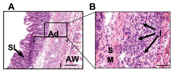
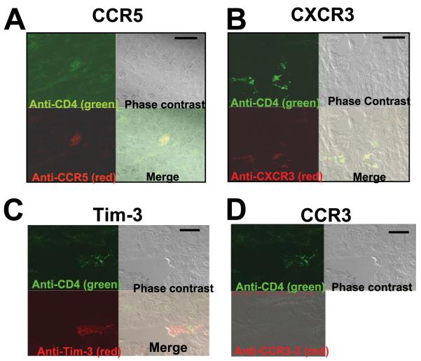
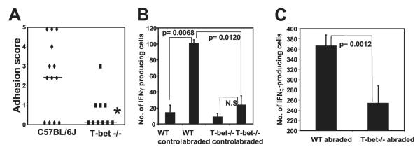
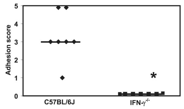
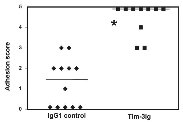
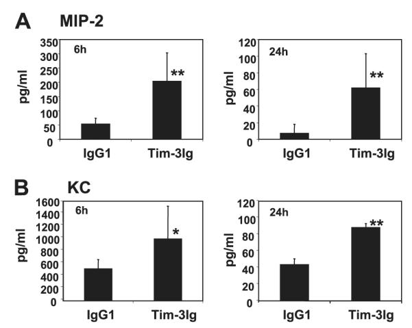
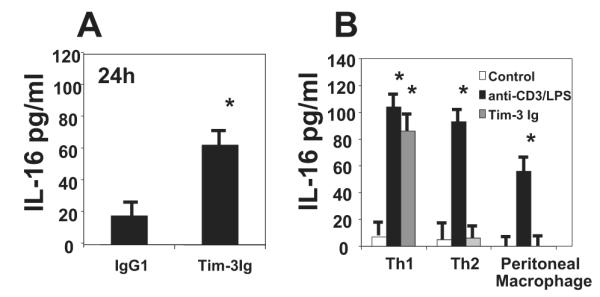
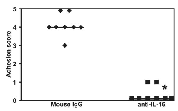
Tissue trauma in the peritoneal and pelvic cavities following surgery or bacterial infection results in adhesions that are a debilitating cause of intestinal obstruction, chronic pelvic pain, and infertility in women. We recently demonstrated that CD4(+) alphabeta T cells are essential for development of this process. Using a murine model of experimental adhesion formation, we now demonstrate that adhesion formation is characterized by the selective recruitment of Tim-3(+), CCR5(+), CXCR3(+), IFN-gamma(+) cells, indicating the presence of a Th1 phenotype. We further demonstrate that adhesion formation is critically dependent on the function of Th1 cells because mice genetically deficient for IFN-gamma, T-bet, or treated with Abs to the Th1-selective chemoattractant IL-16 show significantly less adhesion formation than wild-type mice. In addition, disrupting the interaction of the Th1-specific regulatory molecule Tim-3, with its ligand, significantly exacerbates adhesion formation. This enhanced response is associated with increases in the level of neutrophil-attracting chemokines KC and MIP-2, known to play a role in adhesiogenesis. These data demonstrate that the CD4(+) T cells orchestrating adhesion formation are of the Th1 phenotype and delineate the central role of T-bet, Tim-3, IFN-gamma, and IL-16 in mediating this pathogenic tissue response.
Figures
Similar articles
-
CD4+ T cells regulate surgical and postinfectious adhesion formation.J Exp Med. 2002 Jun 3;195(11):1471-8. doi: 10.1084/jem.20020028. J Exp Med. 2002. PMID: 12045245 Free PMC article.
-
Oral administration with diosgenin enhances the induction of intestinal T helper 1-like regulatory T cells in a murine model of food allergy.Int Immunopharmacol. 2017 Jan;42:59-66. doi: 10.1016/j.intimp.2016.11.021. Epub 2016 Nov 23. Int Immunopharmacol. 2017. PMID: 27886644
-
T-bet is critical for the development of acute graft-versus-host disease through controlling T cell differentiation and function.J Immunol. 2015 Jan 1;194(1):388-97. doi: 10.4049/jimmunol.1401618. Epub 2014 Nov 17. J Immunol. 2015. PMID: 25404360 Free PMC article.
-
Intestinal irradiation and fibrosis in a Th1-deficient environment.Int J Radiat Oncol Biol Phys. 2012 Sep 1;84(1):266-73. doi: 10.1016/j.ijrobp.2011.11.027. Epub 2012 Feb 13. Int J Radiat Oncol Biol Phys. 2012. PMID: 22336200
-
Role of Th1/Th17 balance regulated by T-bet in a mouse model of Mycobacterium avium complex disease.J Immunol. 2014 Feb 15;192(4):1707-17. doi: 10.4049/jimmunol.1302258. Epub 2014 Jan 20. J Immunol. 2014. PMID: 24446514
Cited by
-
An injured tissue affects the opposite intact peritoneum during postoperative adhesion formation.Sci Rep. 2015 Jan 8;5:7668. doi: 10.1038/srep07668. Sci Rep. 2015. PMID: 25566876 Free PMC article.
-
Paricalcitol reduces peritoneal fibrosis in mice through the activation of regulatory T cells and reduction in IL-17 production.PLoS One. 2014 Oct 3;9(10):e108477. doi: 10.1371/journal.pone.0108477. eCollection 2014. PLoS One. 2014. PMID: 25279459 Free PMC article.
-
Comprehensive analysis of chemokines and cytokines secreted in the peritoneal cavity during laparotomy.J Immunoassay Immunochem. 2012;33(3):291-301. doi: 10.1080/15321819.2011.638409. J Immunoassay Immunochem. 2012. PMID: 22738652 Free PMC article.
-
Animal models of intestinal fibrosis: new tools for the understanding of pathogenesis and therapy of human disease.Am J Physiol Gastrointest Liver Physiol. 2012 Oct;303(7):G786-801. doi: 10.1152/ajpgi.00059.2012. Epub 2012 Aug 9. Am J Physiol Gastrointest Liver Physiol. 2012. PMID: 22878121 Free PMC article. Review.
-
Prevention of Post-Operative Adhesions: A Comprehensive Review of Present and Emerging Strategies.Biomolecules. 2021 Jul 14;11(7):1027. doi: 10.3390/biom11071027. Biomolecules. 2021. PMID: 34356652 Free PMC article. Review.
References
-
- Trimbos-Kemper T, Trimbos J, van Hall E. Adhesion formation after tubal surgery: results of the eighth-day laparoscopy in 188 patients. Fertil. Steril. 1985;43:395–400. - PubMed
-
- Monk BJ, Berman M, Montz F. Adhesions after extensive gynecologic surgery: clinical significance, etiology, and prevention. Am. J. Obstet. Gynecol. 1994;170:1396–1403. - PubMed
-
- Lower A, Hawthorn RJ, Ellis H, O’Brien F, Buchan S, Crowe AM. The impact of adhesions on hospital readmissions over ten years after 8849 open gynaecological operations: an assessment from the Surgical and Clinical Adhesions Research Study. Br. J. Obstet. Gynaecol. 2000;107:855–862. - PubMed
-
- Ellis H. The clinical significance of adhesions: focus on intestinal obstruction. Eur. J. Surg. 1997;(Suppl. 577):5–9. - PubMed
-
- Wilson M, Hawkswell J, McCloy RF. Natural history of adhesional small bowel obstruction: counting the cost. Br. J. Surg. 1998;85:1294–1298. - PubMed
Publication types
MeSH terms
Substances
Grants and funding
LinkOut - more resources
Full Text Sources
Other Literature Sources
Medical
Molecular Biology Databases
Research Materials
Miscellaneous
