. 2008;21(5-6):357-72.
doi: 10.1159/000129628. Epub 2008 Apr 24.

KCNQ1 and KCNE1 K+ channel components are involved in early left-right patterning in Xenopus laevis embryos

Affiliations

KCNQ1 and KCNE1 K+ channel components are involved in early left-right patterning in Xenopus laevis embryos

Junji Morokuma et al. Cell Physiol Biochem. 2008.

Abstract

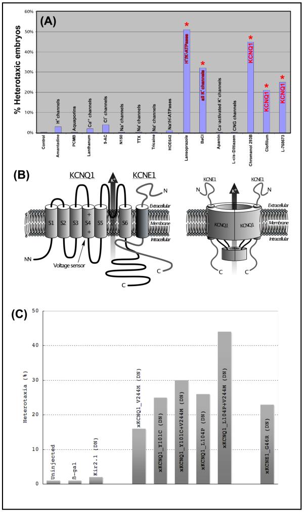
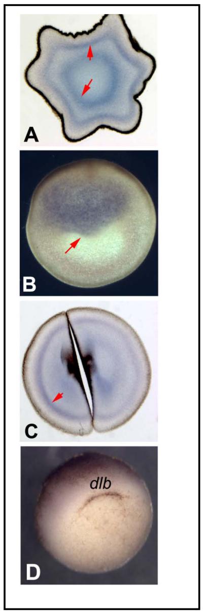
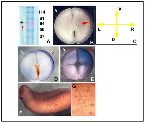
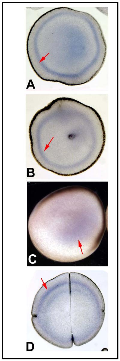
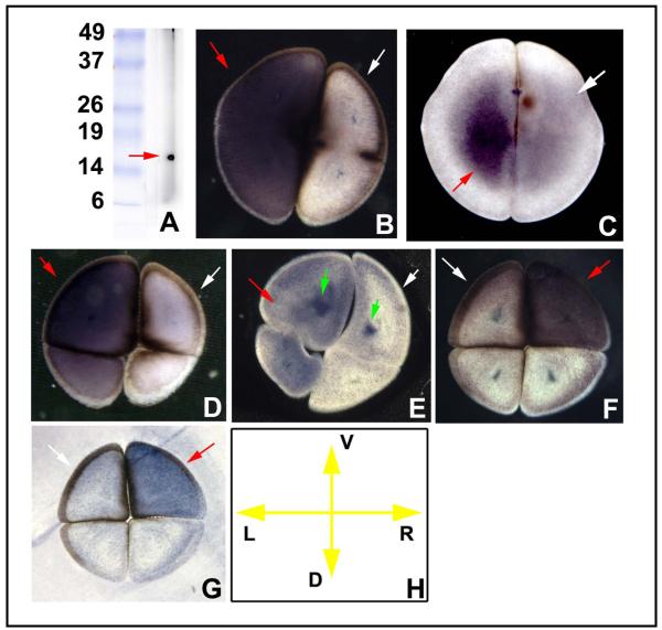
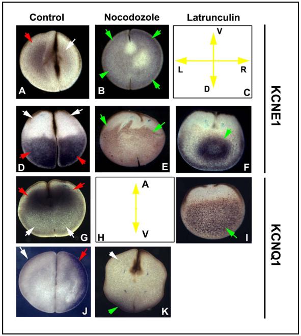
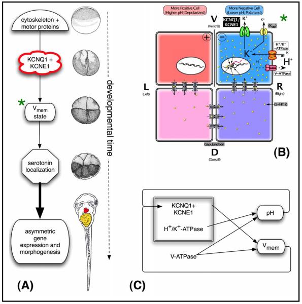
Several ion transporters have been implicated in left-right (LR) patterning. Here, we characterize a new component of the early bioelectrical circuit: the potassium channel KCNQ1 and its accessory subunit KCNE1. Having cloned the native Xenopus versions of both genes, we show that both are asymmetrically localized as maternal proteins during the first few cleavages of frog embryo development in a process dependent on microtubule and actin organization. Molecular loss-of-function using dominant negative constructs demonstrates that both gene products are required for normal LR asymmetry. We propose a model whereby these channels provide an exit path for K(+) ions brought in by the H(+),K(+)-ATPase. This physiological module thus allows the obligate but electroneutral H(+),K(+)-ATPase to generate an asymmetric voltage gradient on the left and right sides. Our data reveal a new, bioelectrical component of the mechanisms patterning a large-scale axis in vertebrate embryogenesis.


Figures

Fig. 1
Fig. 1
A drug screen implicates XKCNQ1 as a high-priority candidate in LR asymmetry. (A) Summary of drug screen data demonstrating that inhibitors targeting XKCNQ1, or the previously-characterized H+/K+-ATPase (positive control), specifically induce significant levels of the independent situs of the heart, gut, and gall-bladder, whereas a variety of other blockers do not. Embryos were exposed to each compound immediately after fertilization and washed out into 0.1X MMR medium at stage 8. They were scored for heterotaxia at st. 44. The doses uses were as follows: HOE642 2.6 mM, spermidine 95 μM, Lanthanum 100 μM, 9-anthracenecarboxylic acid 67 μM, tetrodotoxin 31 μM, MS-222 1.9 μM, Apamin 6.1 μM. The concentrations reported in the literature for these reagents derive largely from the characterization of mammalian channels. Since the Xenopus versions may be somewhat divergent in sequence (and thus have a structure that may not bind the blocker with the same avidity), somewhat greater doses were utilized to reduce the probability of false negatives and to increase the validity of the negative results (which is the case for most of the reagents we explored). (B) KCNQ1 (also known as KvLQT-1) is a 6-transmembrane member of the K+ channel family (schematic modeled on those in [144, 145]). When co-assembled with KCNE1 (also known as minK), it forms the “slow delayed rectifier” or Iks channel [30]. KCNQ1 has different pharmacological profiles depending on whether it is associated with KCNE1 [32, 33]. (C) Histogram summarizing the effects of misexpression of molecular dominant negative constructs (injected at 1-cell stage, and analyzed at st. 44 for situs of heart, gut, and gall-bladder, Table 2A). Microinjections of control mRNAs, including β-gal and a Kir2.1 dominant negative [146], has no significant effect on laterality, while misexpression of dominant negative mutations of XKCNE1 or XKCNQ1 induced significant incidence of heterotaxia. Two of the XKCNQ1 mutations (recovered in human cases of long QT syndrome, labeled as “L104V+V244M”) were additive and induced a higher incidence of laterality phenotype than either mutant alone.
Fig. 2
Fig. 2
Expression of XKCNQ1 mRNA. In situ hybridization was performed on embryos at various stages with an antisense probe to native Xenopus KCNQ1. Panels A and C show JB4 sections made after hybridization in wholemount, while section B shows hybridization made directly on a gelatin-albumin section as described in [44]. (A) Eggs sectioned perpendicularly to the animal-vegetal (AV) axis contain two concentric domains of maternal XKCNQ1 mRNA. (B) Sections made through a fertilized egg along the animal-vegetal axis revealed the presence of maternal mRNA in the animal half of the egg. (C) Embryos at the 2-cell stage sectioned perpendicular to the AV axis reveal maternal mRNA in both blastomeres. (D) Gastrulating embryos show no expression in the organizer or elsewhere.
Fig. 3
Fig. 3
Localization of XKCNQ1 protein. (A) Western blotting of frog embryo extract against the XKCNQ1 antibody revealed a single clean band of the expected size. This antibody was then used on gelatin-albumin sections [44] of early embryos in alkaline-phosphatase immunohistochemistry. Embryos at the 2-cell stage sectioned perpendicular to the AV axis exhibited LR-asymmetric localization at the cell membrane and the immediately adjacent cortex (B). Section orientation is schematized in panel C (V=ventral, D=dorsal, L=left, R=right). At the late 2-cell stage, the stain became more localized to the ventral half of the cell (D) and by the 4-cell stage this could be oriented such that the positive cell was the right ventral one (E). Staining in wholemount the epidermis of the tailbud embryo (F) revealed signal in a subset of the skin cells, which in closeup exhibited the cell-membrane localization (green arrowhead) predicted for an ion channel subunit (G).
Fig. 4
Fig. 4
Expression of XKCNE1 mRNA. Sections through mature oocytes (A) and fertilized eggs (B) both revealed a circumferential pattern of maternal XKCNE1 mRNA. In situ hybridization performed on sections taken through the animal-vegetal axis of fertilized eggs revealed maternal mRNA in the middle portion of the cell (C). During subsequent cleavages, mRNA was detected in the blastomeres in a thin band below the cell cortex (D).
Fig. 5
Fig. 5
Localization of XKCNE1 protein. (A) Western blotting of frog embryo extract against the KCNE1 antibody revealed a single band of the predicted size. Immunoshitochemistry on gelatin-albumin sections taken perpendicular to the AV axis revealed asymmetric staining, which varied among left-handed blastomeres evenly-filled with signal (B) and more vegetal sections which exhibited staining mainly in the center (C), shown to be ventral at the 4-cell stage (D). In more vegetal sections at the 4-cell stage, central spots were observed (E), and in an equal number of embryos, it was the right ventral cell that was positive (F,G). Section orientation is schematized in panel H (V=ventral, D=dorsal, L=left, R=right). Red arrows indicate positive signal; white arrows indicate lack of signal.
Fig. 6
Fig. 6
Localization of XKCNE1 and XKCNQ1 depends on microtubule and actin cytoskeleton. Embryos were treated with disruptors of microtubule organization (Nocodozole) or of actin organization (Latrunculin) for 2 hours after fertilization, and the embryos were fixed, sectioned in different planes, and processed for immunohistochemistry with KCNE1 and XKCNQ1 antibodies. The normal LR-asymmetric localization of XKCNE1 protein is altered from an asymmetric pattern (A) to a symmetrical one when embryos are exposed to Nocodozole (B). Section orientation is schematized in (C) (V=ventral, D=dorsal, L=left, R=right). Along the AV axis, the normal vegetal localization of XKCNE1 (D) is expanded towards the animal pole by disruption of the microtubules (E) and coalesced into a more restricted pattern in the center of the cell by disruption of the actin cytoskeleton (F). Likewise, the normal animal-pole localization of XKCNQ1 (G), schematized in panel H, is converted to a vegetal localization by Latrunculin (I), and the normal LR-asymmetric localization (J) is converted into a dorsal pattern (K) by exposure to Nocodozole. The cleavage furrows in some panels (e.g. F, I, and E) are not apparent even though all embryos are the same age because disruption of cytoskeleton also perturbs cytokinesis. Red arrows indicate positive signal; white arrows indicate lack of signal; green arrows indicate signal that is in an aberrant location.
Fig. 7
Fig. 7
A model of XKCNQ1/XKCNE1 function in LR patterning. (A) Functional steps and a timeline of representative Xenopus stages of a model integrating the data on XKCNQ1/XKCNE1 function into known mechanisms of LR patterning [7, 10]. We propose that the early localization of XKCNQ1 and XKCNE1 is determined by cytoskeleton-dependent intracellular transport, analogously to other LR-relevant ion transporters [15, 77]. The asymmetric localization of this channel complex results in differential ion transport on the Left and Right sides, contributing to membrane voltage stage in ventral blastomeres across the midline [13, 15]. This physiological asymmetry controls downstream laterality pathways through serotonin localization [20, 91] or other transducers such as calcium signaling [21, 92], ultimately leading to the correct morphogenesis of the heart and viscera. Right column shows approximate Xenopus developmental stages corresponding to each event (diagrams taken from [42]). (B) A more detailed model of the module (marked with green asterisk) in panel A, whereby XKCNQ1 contributes to the asymmetries in membrane voltage during early cleavages. Specifically, we propose that this potassium channel (and perhaps others) provides an exit path for the extra K+ ions brought in by the H+/K+-ATPase, thus (analogously to their cooperation in the mammalian gut) allowing this electroneutral pump to generate a voltage gradient by the net loss of positive charges. As we proposed previously [11, 147], asymmetries in XKCNQ1 in ventral cells across the LR midline can contribute to the electrophoretic force responsible for the asymmetric distribution of small molecule signals across gap junctional paths, resulting in asymmetric gene expression. (C) Complex feedback circuits control the bioelectrical state of the early blastomeres. The V-ATPase directly produces a pH gradient and helps set the membrane voltage gradient. The H+/K+-ATPase, in concert with potassium channels like KCNQ1, also contributes to transmembrane potential. Importantly, the KCNQ1/KCNE1 channel is sensitive to both membrane voltage and pH. Thus, this system may exhibit interesting and possibly non-linear dynamics over both the short-term and steady-state time scales, necessitating quantitative modeling in future work.

Similar articles

Cited by

References

    1. McCaig CD, Rajnicek AM, Song B, Zhao M. Controlling cell behavior electrically: Current views and future potential. Physiol Rev. 2005;85:943–978. - PubMed
    1. Levin M. Large-scale biophysics: Ion flows and regeneration. Trends Cell Biol. 2007;17:262–271. - PubMed
    1. Cone CD. Unified theory on the basic mechanism of normal mitotic control and oncogenesis. J Theor Biol. 1971;30:151–181. - PubMed
    1. Levin M. Bioelectromagnetic patterning fields: Roles in embryonic development, regeneration, and neoplasm. Bioelectromagnetics. 2003;24:295–315. - PubMed
    1. Robinson KR, Messerli MA. Left/right, up/down: The role of endogenous electrical fields as directional signals in development, repair and invasion. Bioessays. 2003;25:759–766. - PubMed

Publication types

MeSH terms