. 2008 Jun;40(6):794-9.
doi: 10.1038/ng.126. Epub 2008 May 4.

The mouse X chromosome is enriched for multicopy testis genes showing postmeiotic expression

Affiliations

The mouse X chromosome is enriched for multicopy testis genes showing postmeiotic expression

Jacob L Mueller et al. Nat Genet. 2008 Jun.

Abstract

According to the prevailing view, mammalian X chromosomes are enriched in spermatogenesis genes expressed before meiosis and deficient in spermatogenesis genes expressed after meiosis. The paucity of postmeiotic genes on the X chromosome has been interpreted as a consequence of meiotic sex chromosome inactivation (MSCI)--the complete silencing of genes on the XY bivalent at meiotic prophase. Recent studies have concluded that MSCI-initiated silencing persists beyond meiosis and that most genes on the X chromosome remain repressed in round spermatids. Here, we report that 33 multicopy gene families, representing approximately 273 mouse X-linked genes, are expressed in the testis and that this expression is predominantly in postmeiotic cells. RNA FISH and microarray analysis show that the maintenance of X chromosome postmeiotic repression is incomplete. Furthermore, X-linked multicopy genes exhibit a similar degree of expression as autosomal genes. Thus, not only is the mouse X chromosome enriched for spermatogenesis genes functioning before meiosis, but in addition, approximately 18% of mouse X-linked genes are expressed in postmeiotic cells.


Figures

Figure 1
Figure 1
Mouse X chromosome ampliconic regions containing testis-expressed genes. a, Examples of complexity (Amp1, left), massive scale (Amp4, center), and tandem duplications (Amp19, right) in ampliconic regions. Each ampliconic region is compared to itself in a triangular dot-plot. Individual dots represent a perfect match of 200 nucleotides. Horizontal lines and vertical lines depict direct and inverted repeats, respectively. Shaded grey regions represent physical gaps in the sequence assembly. Primary (black) and secondary (grey) amplicons, or repeat units, are shown below the dot-plots as arrows. The size of each region is shown below the amplicon arrows. b, Mouse X chromosome with ampliconic regions shown in blue. c, Position of ampliconic and non-ampliconic multi-copy genes and their copy number. Asterisks denote copy number estimates since they fall within incompletely assembled genomic regions. Copy number estimates are based upon the number of intact ORFs in the current NCBI Build 37.1.
Figure 2
Figure 2
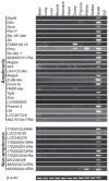
Mouse X chromosome ampliconic and non-ampliconic multi-copy genes exhibit testis-biased expression as shown by RT-PCR. We assayed 26 ampliconic and 10 non-ampliconic multi-copy genes across 11 different tissues – Cxx and Obp1 were not detected in any tissues and are therefore not shown. Of the 35 multi-copy genes expressed in testis, only Xlr4 did not exhibit testis-biased expression. Other Xlr gene family members (Xlr3 and Xlr5) exhibit ubiquitous expression patterns similar to Xlr4 (data not shown). β-Actin is ubiquitously expressed and serves as a control.
Figure 3
Figure 3
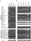
Mouse X chromosome ampliconic and non-ampliconic multi-copy genes are expressed predominantly in germ cells during post-meiotic spermatogenesis as shown by RT-PCR. a. All 23 multi-copy genes exhibiting testis-biased expression were assayed in wild type and germ cell negative XXSry and XXSxrb testes. Only Rhox, PABP-like, Srsx, Pramel-3 and Ott show expression in both XXSry and XXSxrb testes. We note that Pramel-3 gives faint products, consistent with the findings of Wang et al. (2001) that the predominant site of expression for this multi-copy gene is in germ cells. b, Genes exhibiting strict germ cell-specificity were assayed by analysis of the first wave of spermatogenesis in juvenile male mice. Twenty of the 28 genes are expressed from 18.5dpp, the remaining eight – Sstx, Fthl17, Zfp161-like, Magea, ZxdB-like, Dmrtc1b, 170003E24Rik and 1700010D01Rik are expressed from 7.5dpp.
Figure 4
Figure 4
Multi-copy ampliconic genes exhibit higher levels of reactivation in round spermatids than single-copy genes. Four single-copy and three multi-copy X-linked genes were assayed for expression during successive stages of spermatogenesis by RNA/DNA FISH. First column – spermatogonia. All four single-copy genes are expressed at this stage, consistent with previously reported observations (see below for details of quantitation). In contrast, only one multi-copy gene, Ott, is expressed. Second column – pachytene spermatocytes. The sex body is shown by γH2AX staining. All seven genes are silent in 100% of cells examined (n=100). Third and fourth columns represent examples of expressing and non-expressing round spermatid cells, respectively. Single-copy genes show reactivation in a minority of round spermatids, as shown by the percentages in the final column (see also Supplementary Table 4). Birc4 expression could not be detected in any round spermatids, and so the ‘spermatid-expressing’ column is blank for this gene. In contrast, multi-copy genes show expression in a high percentage of round spermatids. Note that although Ott is neither germ cell-specific nor spermatid-specific by RT-PCR, RNA FISH reveals high reactivation levels in round spermatids. For spermatogonial RNA FISH quantitation (n=100 for each gene), cells were substaged based on characteristic cytological criteria. Birc4, Fmr1 and Zfx are expressed in all spermatogonial subtypes, with 97%, 87% and 91% of combined spermatogonia expressing, respectively. Scml2 is expressed in 97% of type A spermatogonia but is silent in other spermatogonial substages. Ott is expressed in 99% of type A spermatogonia and 56% of intermediate and type B spermatogonia.
Figure 5
Figure 5
Microarray analyses of single-copy and multi-copy genes on the X chromosome. a, Mean expression levels (+/− two standard error from mean) of 278 single-copy genes, previously defined as being expressed solely in spermatogonia, in A and B spermatogonia (AS and BS), pachytene spermatocytes (PS) and round spermatids (RS). b, Mean expression levels of autosomal (A), X-linked single-copy (XSC), and X-linked multi-copy (XMC) genes in pachytene spermatocytes and round spermatids. P-values were determined via Wilcoxon Ranks Sum tests. c, Model: Multi-copy genes evade the effects of X chromosome post-meiotic repression. Single-copy and multi-copy X-linked genes exhibit similar average levels of expression during pre-meiotic spermatogenesis. All X-linked genes are subsequently silenced during MSCI. Following MSCI, single-copy X-linked genes exhibit low reactivation levels, fitting with the post-meiotic repression of the X chromosome. Multi-copy X-linked genes exhibit expression levels similar to autosomal genes, thus evading the repressive effects of post-meiotic repression.

Comment in

  • The not-so-silent X.
    Disteche CM. Disteche CM. Nat Genet. 2008 Jun;40(6):689-90. doi: 10.1038/ng0608-689. Nat Genet. 2008. PMID: 18509310 No abstract available.

References

    1. Wang PJ, McCarrey JR, Yang F, Page DC. An abundance of X-linked genes expressed in spermatogonia. Nat Genet. 2001;27:422–6. - PubMed
    1. Khil PP, Smirnova NA, Romanienko PJ, Camerini-Otero RD. The mouse X chromosome is enriched for sex-biased genes not subject to selection by meiotic sex chromosome inactivation. Nat Genet. 2004;36:642–6. - PubMed
    1. Reinke V. Sex and the genome. Nat Genet. 2004;36:548–9. - PubMed
    1. McKee BD, Handel MA. Sex chromosomes, recombination, and chromatin conformation. Chromosoma. 1993;102:71–80. - PubMed
    1. Turner JM. Meiotic sex chromosome inactivation. Development. 2007;134:1823–31. - PubMed

Publication types