. 2008 Jun;9(6):641-9.
doi: 10.1038/ni.1610. Epub 2008 May 4.

The differentiation of human T(H)-17 cells requires transforming growth factor-beta and induction of the nuclear receptor RORgammat

Affiliations

The differentiation of human T(H)-17 cells requires transforming growth factor-beta and induction of the nuclear receptor RORgammat

Nicolas Manel et al. Nat Immunol. 2008 Jun.

Abstract

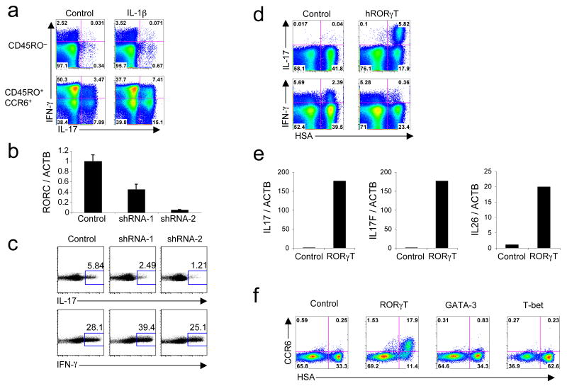
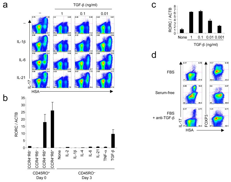
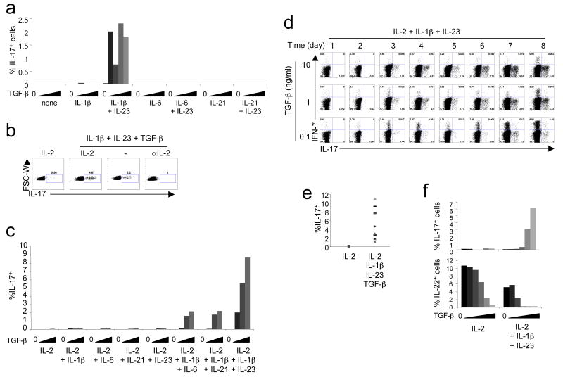
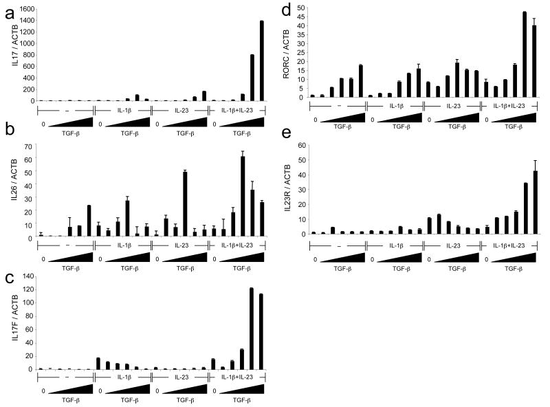
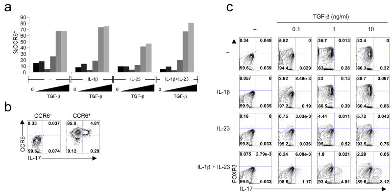
T(H)-17 cells are interleukin 17 (IL-17)-secreting CD4+ T helper cells involved in autoimmune disease and mucosal immunity. In naive CD4+ T cells from mice, IL-17 is expressed in response to a combination of IL-6 or IL-21 and transforming growth factor-beta (TGF-beta) and requires induction of the nuclear receptor RORgammat. It has been suggested that the differentiation of human T(H)-17 cells is independent of TGF-beta and thus differs fundamentally from that in mice. We show here that TGF-beta, IL-1beta and IL-6, IL-21 or IL-23 in serum-free conditions were necessary and sufficient to induce IL-17 expression in naive human CD4+ T cells from cord blood. TGF-beta upregulated RORgammat expression but simultaneously inhibited its ability to induce IL-17 expression. Inflammatory cytokines relieved this inhibition and increased RORgammat-directed IL-17 expression. Other gene products detected in T(H)-17 cells after RORgammat induction included the chemokine receptor CCR6, the IL-23 receptor, IL-17F and IL-26. Our studies identify RORgammat as having a central function in the differentiation of human T(H)-17 cells from naive CD4+ T cells and suggest that similar cytokine pathways are involved in this process in mice and humans.


Figures

Figure 1
Figure 1. RORγT is necessary and sufficient for the expression of IL-17 in human CD4+ T cells
(a) Flow cytometry on sorted CD45RO and CD45RO+CCR6+ activated and expanded in the presence of IL-2 with or without IL-1β. IL-17 and IFN-γ production was analyzed at day 6. (b,c) RT-PCR for RORC and ACTB mRNA expression (b) and flow cytometry for intracellular IL-17 and IFN-γ (c) in sorted CD45RO+CCR6+ cells transduced with an empty vector or vector encoding for RORγT-specific shRNA (shRNA-1 and shRNA-2). Cells were selected in puromycin at day 2 and expression of mRNA and cytokines was analyzed at day 6. Data are representative of four independent experiments. (d) Flow cytometry of naive cord blood CD4+ T cells activated, transduced by vectors encoding IRES-HSA or RORγT-IRES-HSA and then expanded for 6 days in the presence of IL-2. Intracellular IL-17 and IFN-γ production was analyzed at day 6. (e) RT-PCR of ACTB, IL17, IL17F and IL26 in naive cord blood CD4+ T cells transduced with vectors encoding IRES-GFP or RORγT-IRES-GFP. GFP+ cells were sorted at day 6 and the levels of mRNAs were analyzed. (f) Flow cytometry of naive cord blood CD4+ T cells transduced with vectors encoding IRES-HSA, RORγT-IRES-HSA, GATA-3-IRES-HSA or T-bet-IRES-HSA. CCR6 cell surface expression was measured at day 12. Each panel is representative of three independent experiments unless noted otherwise.
Figure 2
Figure 2. TGF-β induces RORγT and inhibits its activity, but the inhibition is relieved by inflammatory cytokines
(a) Flow cytometry of naive cord blood CD4+ T cells transduced with a vector encoding RORγT-IRES-HSA alone or with combination of IL-1β, IL-6, IL-21 and increasing concentrations of TGF-β. IL-17 intracellular staining was performed at day 6. (b,c) RT-PCR of RORC and ACTB expression measured in freshly sorted CCR4−/+CCR6−/+ adult memory CD4+ T cells and in naive CD4+ T cells cultivated for 3 days in the presence of various cytokines (b) and in naive cord blood CD4+ T cells cultivated with various concentrations of TGF-β (c) (d) Flow cytometry of naive cord blood CD4+ T cells transduced with a vector encoding RORγT-IRES-HSA in serum-containing medium with or without anti-TGF-β or in serum-free medium. Intracellular IL-17 and FOXP3 expression was analyzed at day 6. A representative donor with low IL-17 expression following RORγT transduction in the presence of serum is shown. Each panel is representative of at least three independent experiments.
Figure 3
Figure 3. TGF-β, IL-1β and IL-6, IL-21 or IL-23 are required for human TH-17 cell polarization in serum-free conditions
(a) Naive cord blood CD4+ T cells were activated without cytokines or with IL-1β, IL-6 or IL-21 with or without IL-23, alone or with increasing concentrations of TGF-β. IL-2 was added at day 3 and IL-17 expression was analyzed at day 14. (b) Naive cord blood CD4+ T cells were activated with no cytokine or with a combination of IL-1β + IL-23 + TGF-β (1 ng/ml), with or without 10 U/ml IL-2 or neutralizing anti-IL-2. IL-17 expression was analyzed at day 6. (c) Naive cord blood CD4+ T cells were cultivated with IL-2 and different concentrations of TGF-β in the presence of the listed cytokines. IL-17 expression was analyzed at day 6. (d) Time-course of IL-17 and IFN-γ production in naive cord blood CD4+ T cells polarized in the presence of IL-2 + IL-1β + IL-23 + TGF-β (0.1, 1 or 10 ng/ml). (e) Summary of IL-17 expression in day 6 cultures of naive cord blood CD4+ T cells from different donors (n = 11) in the presence of IL-2 + IL-1β + IL-23 + 10 ng/ml TGF-β. (f) Naive cord blood CD4+ T cells were cultivated in IL-2 alone or IL-23 + IL-1β + IL-2 with increasing concentrations of TGF-β. IL-17 and IL-22 expression were analyzed at day 6. Each panel is representative of at least three independent donors.
Figure 4
Figure 4. Induction of IL26, IL17F, IL17, RORC and IL23R mRNAs during human TH-17 cell differentiation
(a-c) Naive cord blood CD4+ T cells were cultivated with IL-2 alone or with IL-1β + IL-2, IL-23 + IL-2 or IL-23 + IL-1β + IL-2 with increasing concentrations of TGF-β. mRNA levels of ACTB, IL17 (a), IL26 (b) and IL17F (c) were analyzed on day 6 following restimulation with PMA and ionomycin. (d, e) Naive cord blood CD4+ T cells were cultivated with IL-2 alone or with IL-1β + IL-2, IL-23 + IL-2 or IL-23 + IL-1β + IL-2 with increasing concentrations of TGF-β. mRNA levels of ACTB, RORC (d) and IL23R (e) were analyzed on day 6. Each panel is representative of three independent donors.
Figure 5
Figure 5. Expression of CCR6 and FOXP3 during human TH-17 cell differentiation
(a) Naive cord blood CD4+ T cells were cultivated with IL-2 alone or with IL-1β + IL-2, IL-23 + IL-2 or IL-23 + IL-1β + IL-2 with increasing concentrations of TGF-β. CCR6 expression was analyzed by surface staining at day 6. (b) Naive cord blood CD4+ T cells were cultivated for 6 days in IL-2 + IL-23 + IL-1β + IL-2 + 10 ng/ml TGF-β. CCR6+ and CCR6- cells were sorted and intracellular IL-17 was analyzed. (c) Naive cord blood CD4+ T cells were cultivated with IL-2 alone or with IL-1β + IL-2, IL-23 + IL-2 or IL-23 + IL-1β + IL-2 with increasing concentrations of TGF-β. FOXP3 and IL-17 expression were analyzed at day 6. Data are representative of four independent experiments.

Comment in

References

    1. Weaver CT, Hatton RD, Mangan PR, Harrington LE. IL-17 family cytokines and the expanding diversity of effector T cell lineages. Annual review of immunology. 2007;25:821–852. - PubMed
    1. Bettelli E, Korn T, Kuchroo VK. Th17: the third member of the effector T cell trilogy. Current opinion in immunology. 2007;19:652–657. - PMC - PubMed
    1. Stockinger B, Veldhoen M. Differentiation and function of Th17 T cells. Current opinion in immunology. 2007;19:281–286. - PubMed
    1. Acosta-Rodriguez EV, et al. Surface phenotype and antigenic specificity of human interleukin 17-producing T helper memory cells. Nature immunology. 2007;8:639–646. - PubMed
    1. Annunziato F, et al. Phenotypic and functional features of human Th17 cells. J Exp Med. 2007;204:1849–1861. - PMC - PubMed

Publication types

MeSH terms

Substances