. 2008 Jun;5(6):545-51.
doi: 10.1038/nmeth.1209. Epub 2008 May 4.

Improving the photostability of bright monomeric orange and red fluorescent proteins

Affiliations

Improving the photostability of bright monomeric orange and red fluorescent proteins

Nathan C Shaner et al. Nat Methods. 2008 Jun.

Abstract

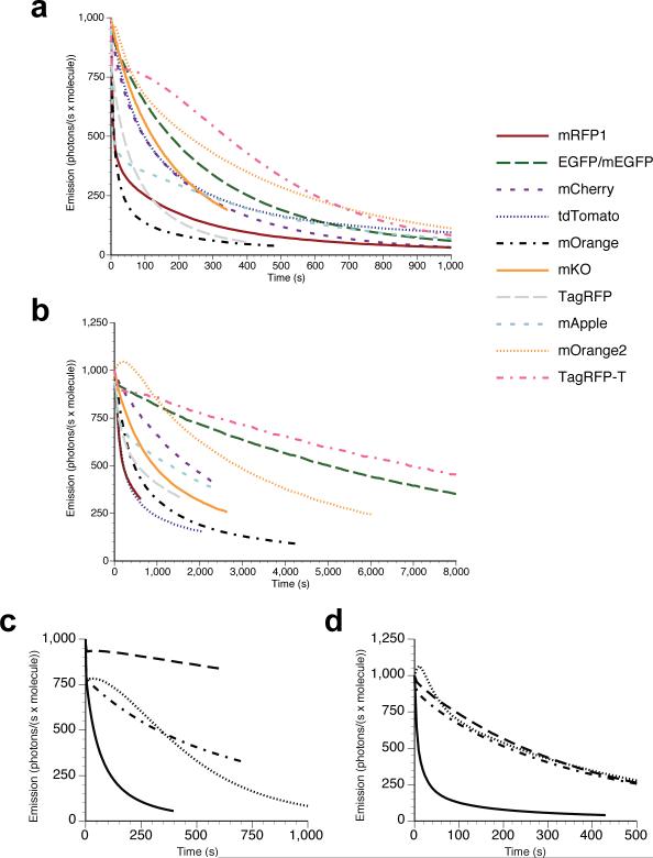
All organic fluorophores undergo irreversible photobleaching during prolonged illumination. Although fluorescent proteins typically bleach at a substantially slower rate than many small-molecule dyes, in many cases the lack of sufficient photostability remains an important limiting factor for experiments requiring large numbers of images of single cells. Screening methods focusing solely on brightness or wavelength are highly effective in optimizing both properties, but the absence of selective pressure for photostability in such screens leads to unpredictable photobleaching behavior in the resulting fluorescent proteins. Here we describe an assay for screening libraries of fluorescent proteins for enhanced photostability. With this assay, we developed highly photostable variants of mOrange (a wavelength-shifted monomeric derivative of DsRed from Discosoma sp.) and TagRFP (a monomeric derivative of eqFP578 from Entacmaea quadricolor) that maintain most of the beneficial qualities of the original proteins and perform as reliably as Aequorea victoria GFP derivatives in fusion constructs.


Figures

Figure 1
Figure 1. Comparison of photobleaching curves
(a) Arc lamp photobleaching curves for mRFP1, EGFP, mCherry, tdTomato, mOrange, mKO, TagRFP, mApple, mOrange2, and TagRFP-T, as measured in purified protein (see Methods) and plotted as intensity versus normalized total exposure time with an initial emission rate of 1000 photons/s per molecule; (b) normalized laser scanning confocal microscopy bleaching curves for the same proteins (except for EGFP which in this case is the monomeric A206K variant) fused to histone 2B and imaged in live cells. The time axis represents normalized total imaging time for an initial scan-averaged emission rate of 1000 photons/s per molecule; (c) arc lamp photobleaching curves for normoxic TagRFP (solid line) and TagRFP-T (dotted line) and O2-free TagRFP (dot-dashed line) and TagRFP-T (dashed line). All photobleaching curves were measured under continuous illumination without neutral density filters and are plotted as intensity versus normalized total exposure time with an initial emission rate of 1000 photons/s per molecule; (d) arc lamp photobleaching curves for normoxic mOrange (solid lines) and mOrange2 (dotted lines) and (O2-free) mOrange (dot-dashed line) and mOrange2 (dashed line).
Figure 2
Figure 2. Fluorescence imaging of TagRFP-T subcellular targeting fusions
N-terminal fusion constructs (linker amino acid length indicated after fusion protein name): (a) TagRFP-T-N1 (N-fusion cloning vector; expression in nucleus and cytoplasm with no specific localization); (b) TagRFP-T-mitochondria-7 (human cytochrome C oxidase subunit VIII); (c) TagRFP-T-H2B-6 (N-terminus; human, showing two interphase nuclei and one nucleus in early anaphase); (d) TagRFP-T-Golgi-7 (N-terminal 81 amino acids of human β-1,4-galactosyltransferase); (e) TagRFP-T-vimentin-7 (human); (f) TagRFP-T-Cx43-7 (rat α-1 connexin-43); (g) TagRFP-T-zyxin-7 (human); C-terminal fusion constructs: (h) TagRFP-T-annexin (A4)-12 (human; illustrated with ionomycin-induced translocation to the plasma and nuclear membranes); (i) TagRFP-T-lamin B1-10 (human); (j) TagRFP-T-vinculin-23 (human); (k) TagRFP-T-clathrin light chain-15 (human); (l) TagRFP-T-β-actin-7 (human); (m) TagRFP-T-peroxisomes-2 (peroximal targeting signal 1; PTS1); (n) TagRFP-T-endosomes-15 (human RhoB GTPase with an N-terminal c-Myc epitope tag); (o) TagRFP-T-farnesyl-5 (20-amino acid farnesylation signal from c-Ha-Ras); (p) TagRFP-T-β-tubulin-6 (human). All TagRFP-T fusion vectors were expressed in HeLa (ATCC; CCL-2) cells. Scale bars are 10 μm.
Figure 3
Figure 3. Fluorescence imaging of mOrange2 subcellular targeting fusions
Widefield fluorescence images of mOrange2 chimeras in N- and C-terminal fusions. N-terminal fusion constructs (linker amino acid length indicated after fusion protein name): (a) mOrange2-Keratin-17 (human cytokeratin 18); (b) mOrange2-Cx26-7 (rat β-2 connexin-26); (c) mOrange2-Golgi-7 (N-terminal 81 amino acids of human β-1,4-galactosyltransferase); (d) mOrange2-vimentin-7 (human); (e) mOrange2-EB3-7 (human microtubule-associated protein; RP/EB family); (f) mOrange2-mitochondria-7 (human cytochrome C oxidase subunit VIII); (g) mOrange2-paxillin-22 (chicken); (h) mOrange2-α-actinin-19 (human non-muscle); C-terminal fusion constructs: (i) mOrange2-Lamin B1-10 (human); (j) mOrange2-β-Actin-7 (human); (k) mOrange2-lysosomes-20 (rat lysosomal membrane glycoprotein 1); (l) mOrange2-peroxisomes-2 (peroximal targeting signal 1); (m) mOrange2-β-tubulin-6 (human); (n) mOrange2-Fibrillarin-7 (human); (o) mOrange2-vinculin-23 (human); (p) mOrange2-Clathrin Light Chain-15 (human). (qu) Laser scanning confocal images of HeLa cells expressing mOrange2-H2B-6 (N-terminal fusion; human) progressing through (q) interphase; (r) prophase; (s) prometaphase; (t) metaphase; (u) early anaphase. The cell line used for expressing mOrange2 fusion vectors was Gray fox lung fibroblast cells (FoLu) in panels (e) and (j), and human cervical adenocarcinoma cells (HeLa) in the remaining panels. Scale bars are 10 μm.

Comment in

References

    1. Campbell RE, et al. A monomeric red fluorescent protein. Proc Natl Acad Sci U S A. 2002;99:7877–7882. - PMC - PubMed
    1. Shaner NC, et al. Improved monomeric red, orange and yellow fluorescent proteins derived from Discosoma sp. red fluorescent protein. Nat Biotechnol. 2004;22:1567–1572. - PubMed
    1. Chudakov DM, et al. Photoswitchable cyan fluorescent protein for protein tracking. Nat Biotechnol. 2004;22:1435–1439. - PubMed
    1. Griesbeck O, Baird GS, Campbell RE, Zacharias DA, Tsien RY. Reducing the environmental sensitivity of yellow fluorescent protein. Mechanism and applications. J Biol Chem. 2001;276:29188–29194. - PubMed
    1. Habuchi S, et al. Reversible single-molecule photoswitching in the GFP-like fluorescent protein Dronpa. Proc Natl Acad Sci U S A. 2005;102:9511–9516. - PMC - PubMed

Publication types

Associated data