. 2008 Feb 29;4(2):e1000015.
doi: 10.1371/journal.pgen.1000015.

DNA damage activates the SAC in an ATM/ATR-dependent manner, independently of the kinetochore

Affiliations

DNA damage activates the SAC in an ATM/ATR-dependent manner, independently of the kinetochore

Eun Mi Kim et al. PLoS Genet. .

Abstract

The DNA damage checkpoint and the spindle assembly checkpoint (SAC) are two important regulatory mechanisms that respond to different lesions. The DNA damage checkpoint detects DNA damage, initiates protein kinase cascades, and inhibits the cell cycle. The SAC relies on kinetochore-dependent assembly of protein complexes to inhibit mitosis when chromosomes are detached from the spindle. The two checkpoints are thought to function independently. Here we show that yeast cells lacking the DNA damage checkpoint arrest prior to anaphase in response to low doses of the DNA damaging agent methyl methane sulfonate (MMS). The arrest requires the SAC proteins Mad1, Mad2, Mad3, Bub1, and Bub3 and works through Cdc20 and Pds1 but unlike the normal SAC, does not require a functional kinetochore. Mec1 (ATR) and Tel1 (ATM) are also required, independently of Chk1 and Rad53, suggesting that Mec1 and Tel1 inhibit anaphase in response to DNA damage by utilizing SAC proteins. Our results demonstrate cross-talk between the two checkpoints and suggest that assembling inhibitory complexes of SAC proteins at unattached kinetochores is not obligatory for their inhibitory activity. Furthermore, our results suggest that there are novel, important targets of ATM and ATR for cell cycle regulation.


Conflict of interest statement

The authors have declared that no competing interests exist.

Figures
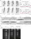
Figure 1
Figure 1. Mad2 is required to delay of mitosis in response to MMS.
(A) Flow cytometry of wild type (WT) and mutant cells with the indicated genotypes that were arrested with α-factor and released into YPD medium in the presence or absence of 0.01% MMS. Cells were assayed every fifteen minutes. The rad9 rad24 and mad2 strains were tested twice, WT was tested six times, and rad9 rad24 mad2 was tested three times. (B) The percentage of large budded bi-nucleate cells (anaphase) from panel A for wild type and indicated mutant cells after release into medium with or without MMS. At least 100 cells were counted for each time point. Cell morphologies indicative of other phases of the cell cycle are in Figure S2. (C) Pds1-13 Myc stability of wild type and mutants cells. Endogenous Pds1 was tagged with 13 copies of the Myc epitope. Protein extracts from the cells in (A) were prepared and Western blot analysis was performed with 9E10 mouse anti-Myc monoclonal antibody. Upper half was in the absence of MMS and lower half was in the presence of MMS. Western blots with anti-Tub2 (tubulin) were for loading control. (D) Wild type and cells of the indicated genotypes were arrested with α-factor and released into YPD medium with or without 0.01% MMS. HU indicates wild type cells arrested with 0.1 M hydroxyurea. Samples were taken every hour and chromosomes were separated by CHEF.
Figure 2
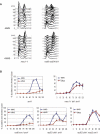
Figure 2. The SAC-dependent mitotic delay is independent of the kinetochore.
(A) Flow cytometry of mutant cells with the indicated genotypes that were arrested with α-factor and released into YPD medium with or without 0.01% MMS. Cells were assayed every fifteen minutes. The rad9 rad24 ndc10-1 cells were arrested with α-factor at 23°C for 3 hours, moved to 35°C for 1 hour to inactivate Ndc10, and then released to pre-warmed YPD at 35°C with or without MMS. All strains were tested twice except rad9 rad24 bub1 which was tested 3 times. (B) The percentage of large budded bi-nucleate cells from panel A for wild type and indicated mutant cells after released into medium without MMS (upper panel) or with MMS (lower panel). Cell morphologies indicative of other phases of the cell cycle are in Figure S4. (C) The number of rad9 rad24 bub3 cells that were budded with divided nuclei (anaphase) when grown in the presence or absence of MMS. Data from two independent experiments are represented. Solid lines are mean values and the dots are the independent measurements (range). (D) The SAC delay in response DNA damage requires APCCdc20. The number of CDC20-127 cells (upper panel) and CDC20-127 rad9 rad24 cells (lower panel) that were budded with divided nuclei (anaphase) when grown in the presence or absence of MMS. Data from three independent experiments are represented. The means are plotted and standard deviation is indicated by error bars. Analyses of morphologies indicative of other phases of cell cycle are in Figure S5D. (E) The SAC delay in response DNA damage requires Pds1. Number of cells that were budded with divided nuclei (anaphase) when grown in the presence of MMS are graphed. Closed circles are rad9 rad24 cells, open circles are rad9 rad24 mad2 cells, and triangles are rad9 rad24 pds1 cells. The arrows represent the time when 50% of the cells had completed anaphase when grown in the absence of MMS. Each point is the mean value of two independent experiments.
Figure 3
Figure 3. Mec1 and Tel1 activate the SAC in response to MMS.
(A) Flow cytometry of mec1-1 and rad9 rad24 tel1 cells arrested with α-factor and released to the cell cycle with or without MMS. (B) The percentage of cells that completed anaphase of isogenic sml1Δ cells (EKY490) and mutants of the indicated genotypes that were arrested with α-factor and had formed mating projections. The mean numbers of cells are graphed for cells with the indicated genotypes and the standard deviations are represented by error bars. Experiments were repeated independently three times and at least one hundred cells were counted for each time point in each experiment.
Figure 4
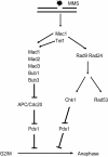
Figure 4. A model for the role of the SAC in response to DNA damage.
The lesion (indicated by the star) activates Mec1 and Tel1 which inhibits anaphase by phosphorylating Pds1 through the DNA damage checkpoint and independently inhibits Pds1 turnover by inhibiting APCCdc20 through the activity of the SAC.

References

    1. Zhou BB, Elledge SJ. The DNA damage response: putting checkpoints in perspective. Nature. 2000;408:433–439. - PubMed
    1. Harrison JC, Haber JE. Surviving the breakup: the DNA damage checkpoint. Annu Rev Genet. 2006;40:209–235. - PubMed
    1. Nyberg KA, Michelson RJ, Putnam CW, Weinert TA. Toward maintaining the genome: DNA damage and replication checkpoints. Annu Rev Genet. 2002;36:617–656. - PubMed
    1. Kastan MB, Bartek J. Cell-cycle checkpoints and cancer. Nature. 2004;432:316–323. - PubMed
    1. Bashkirov VI, King JS, Bashkirova EV, Schmuckli-Maurer J, Heyer WD. DNA repair protein Rad55 is a terminal substrate of the DNA damage checkpoints. Mol Cell Biol. 2000;20:4393–4404. - PMC - PubMed

Publication types

MeSH terms