

Radiosensitization of melanoma cells through combined inhibition of protein regulators of cell survival
- PMID: 18454317
- PMCID: PMC4374428
- DOI: 10.1007/s10495-008-0212-y
Radiosensitization of melanoma cells through combined inhibition of protein regulators of cell survival
Abstract
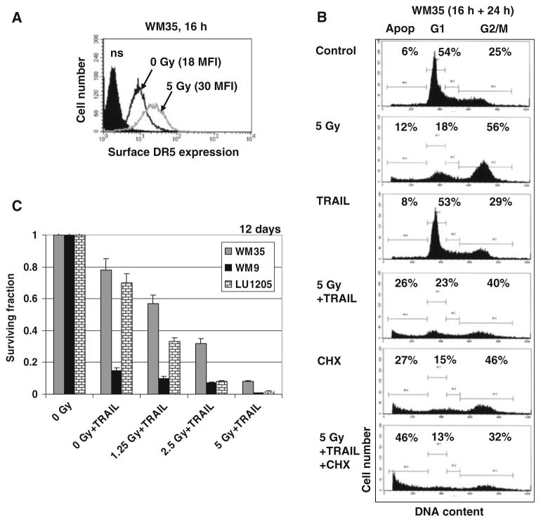
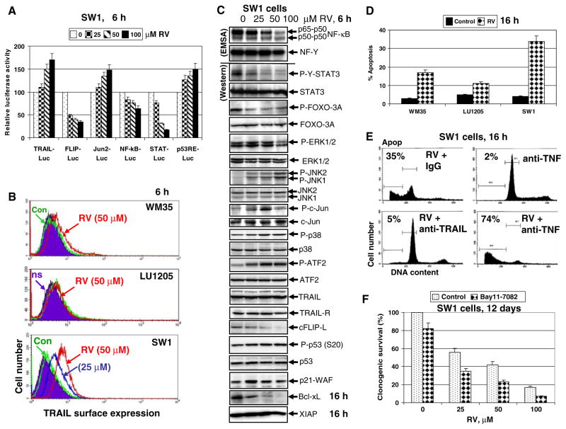
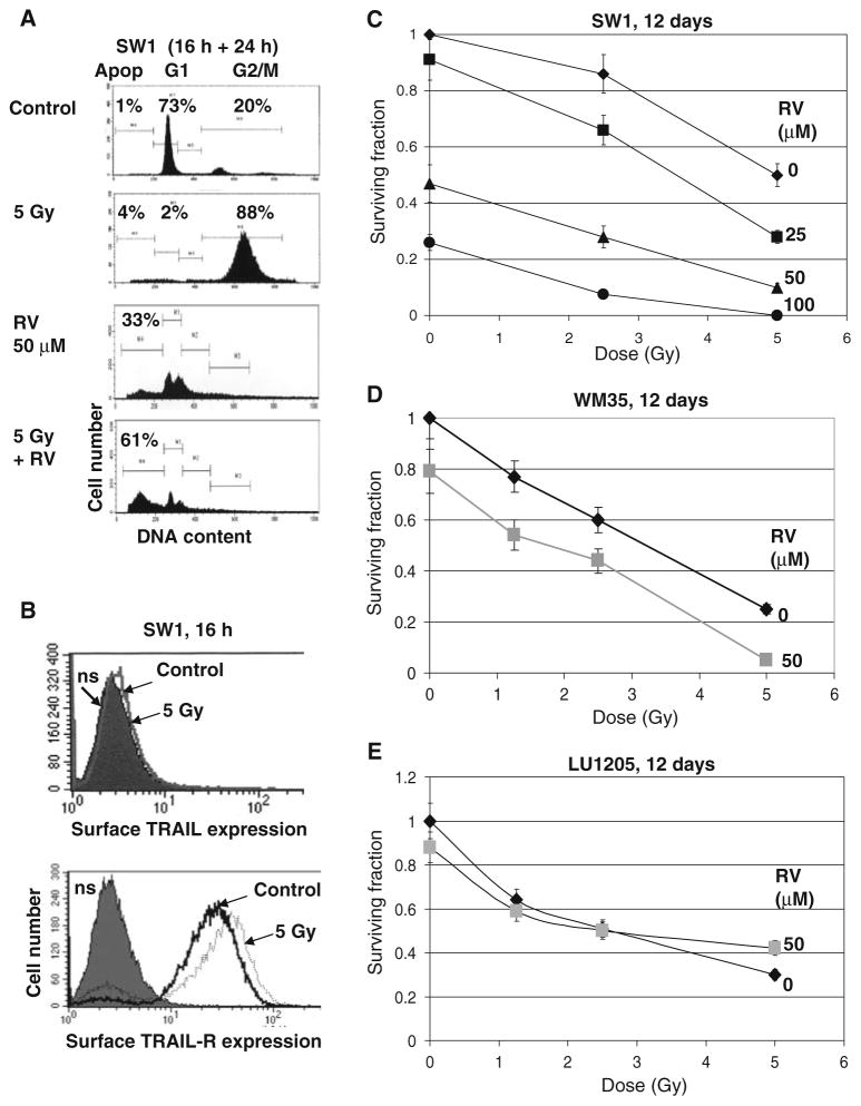
The incidence of melanoma continues to dramatically increase in most Western countries with predominantly Caucasian populations. However, only limited therapies for the metastatic stage of the disease are currently available. The main purpose of this study is to determine approaches that can substantially increase radiosensitivity of melanoma cells. The PI3K-AKT, NF-kappaB and COX-2 pathways, which are involved in the radioprotective response, are highly active in melanoma cells. Pharmacological suppression of COX-2 and PI3K-AKT, or RNAi-mediated knockdown of COX-2, substantially increased levels of G2/M arrest of the cell cycle and decreased clonogenic survival of gamma-irradiated melanomas, predominantly via a necrotic mechanism. On the other hand, resveratrol, a polyphenolic phytoalexin, selectively targets numerous cell signaling pathways, decreasing clonogenic survival primarily via an apoptotic mechanism. In melanoma cells, resveratrol inhibits STAT3 and NF-kappaB-dependent transcription, culminating in suppression of cFLIP and Bcl-xL expression, while activating the MAPK- and the ATM-Chk2-p53 pathways. Resveratrol also upregulates TRAIL promoter activity and induces TRAIL surface expression in some melanoma cell lines, resulting in a rapid development of apoptosis. Sequential treatment of melanoma cells, first with gamma-irradiation to upregulate TRAIL-R surface expression, and then with resveratrol to suppress antiapoptotic proteins cFLIP and Bcl-xL and induce TRAIL surface expression, had dramatic effects on upregulation of apoptosis in some melanoma lines, including SW1 and WM35. However, for melanoma lines exhibiting suppressed translocation of TRAIL to the cell surface, a necrotic mechanism of cell death was primarily involved in radiation response. Hence, surface expression of TRAIL induced by resveratrol appears to be a decisive event, one which determines an apoptotic versus a necrotic response of melanoma cells to sequential treatment.
Figures
Similar articles
-
Resveratrol sensitizes melanomas to TRAIL through modulation of antiapoptotic gene expression.Exp Cell Res. 2008 Mar 10;314(5):1163-76. doi: 10.1016/j.yexcr.2007.12.012. Epub 2007 Dec 23. Exp Cell Res. 2008. PMID: 18222423 Free PMC article.
-
Inhibition of ataxia telangiectasia mutated kinase activity enhances TRAIL-mediated apoptosis in human melanoma cells.Cancer Res. 2009 Apr 15;69(8):3510-9. doi: 10.1158/0008-5472.CAN-08-3883. Epub 2009 Apr 7. Cancer Res. 2009. PMID: 19351839 Free PMC article.
-
Sequential treatment by ionizing radiation and sodium arsenite dramatically accelerates TRAIL-mediated apoptosis of human melanoma cells.Cancer Res. 2007 Jun 1;67(11):5397-407. doi: 10.1158/0008-5472.CAN-07-0551. Cancer Res. 2007. PMID: 17545621 Free PMC article.
-
Resveratrol addiction: to die or not to die.Mol Nutr Food Res. 2009 Jan;53(1):115-28. doi: 10.1002/mnfr.200800148. Mol Nutr Food Res. 2009. PMID: 19072742 Review.
-
Resveratrol modulates phorbol ester-induced pro-inflammatory signal transduction pathways in mouse skin in vivo: NF-kappaB and AP-1 as prime targets.Biochem Pharmacol. 2006 Nov 30;72(11):1506-15. doi: 10.1016/j.bcp.2006.08.005. Epub 2006 Sep 26. Biochem Pharmacol. 2006. PMID: 16999939 Review.
Cited by
-
Therapeutic implications of targeting the PI3Kinase/AKT/mTOR signaling module in melanoma therapy.Am J Cancer Res. 2012;2(2):178-91. Epub 2012 Feb 15. Am J Cancer Res. 2012. PMID: 22485197 Free PMC article.
-
The paradox role of caspase cascade in ionizing radiation therapy.J Biomed Sci. 2016 Dec 7;23(1):88. doi: 10.1186/s12929-016-0306-8. J Biomed Sci. 2016. PMID: 27923354 Free PMC article. Review.
-
Breast cancer cells in three-dimensional culture display an enhanced radioresponse after coordinate targeting of integrin alpha5beta1 and fibronectin.Cancer Res. 2010 Jul 1;70(13):5238-48. doi: 10.1158/0008-5472.CAN-09-2319. Epub 2010 Jun 1. Cancer Res. 2010. PMID: 20516121 Free PMC article.
-
Multifaceted approach to resveratrol bioactivity: Focus on antioxidant action, cell signaling and safety.Oxid Med Cell Longev. 2010 Mar-Apr;3(2):86-100. doi: 10.4161/oxim.3.2.11147. Oxid Med Cell Longev. 2010. PMID: 20716933 Free PMC article. Review.
-
The effect of resveratrol in combination with irradiation and chemotherapy: study using Merkel cell carcinoma cell lines.Strahlenther Onkol. 2014 Jan;190(1):75-80. doi: 10.1007/s00066-013-0445-8. Epub 2013 Nov 8. Strahlenther Onkol. 2014. PMID: 24196280
References
-
- Debatin KM, Krammer PH. Death receptors in chemotherapy and cancer. Oncogene. 2004;23:2950–2966. - PubMed
-
- Okada H, Mak TW. Pathways of apoptotic and non-apoptotic death in tumour cells. Nat Rev Cancer. 2004;4:592–603. - PubMed
-
- Cory S, Adams JM. Killing cancer cells by flipping the Bcl-2/Bax switch. Cancer Cell. 2005;8:5–6. - PubMed
-
- Karin M. Nuclear factor-kappaB in cancer development and progression. Nature. 2006;441:431–436. - PubMed
-
- Krammer PH, Kaminski M, Kiessling M, Gulow K. No life without death. Adv Cancer Res. 2007;97C:111–138. - PubMed
Publication types
MeSH terms
Substances
Grants and funding
LinkOut - more resources
Full Text Sources
Other Literature Sources
Medical
Research Materials
Miscellaneous
