. 2008 Jun;47(6):1994-2002.
doi: 10.1002/hep.22218.

The hepatic stem cell niche: identification by label-retaining cell assay

Affiliations

The hepatic stem cell niche: identification by label-retaining cell assay

Reiichiro Kuwahara et al. Hepatology. 2008 Jun.

Abstract

Label retention assays remain the state-of-the-art approach to identify the location of intraorgan epithelial stem cell niches, in situ and in vivo. They are commonly used in organs with rapid cell turnover but have not been applied to the liver, where cell turnover is very slow. We used a sublethal dose of acetaminophen administered coincident with bromodeoxyuridine to load possible hepatic stem cells in mice with label and then administered a second, sublethal chase of acetaminophen to accomplish "washout" of label from transit amplifying cell populations.

Conclusion: Four possible hepatic stem cell niches are identified by this approach: the canal of Hering (proximal biliary tree), intralobular bile ducts, periductal "null" mononuclear cells, and peribiliary hepatocytes. These results confirm several different and often contradictory lines of investigation regarding the intrahepatic location of stem/progenitor cells and suggest that the liver has a multi-tiered, flexible system of regeneration rather than a single stem/progenitor cell location.


Conflict of interest statement

Potential conflict of interest: Nothing to report.

Figures

Fig. 1
Fig. 1
Summary of experimental procedures for the “label-retaining” cell assay. Mice were fasted for 8 hours before APAP administration, then received intraperitoneal injection of APAP at the dose of 500 mg/kg at day −14, and 750 mg/kg APAP injection at day 0. Water containing BrdU was given before and after APAP administration. Three mice each were sacrificed at days 0, 28, and 56 to perform experiments. BrdU, bromodeoxyuridine; APAP, acetaminophen.
Fig. 2
Fig. 2
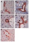
Localization of BrdU-labeled cells in APAP-injured liver. APAP-treated BrdU-labeled murine liver was double-immunostained for BrdU (brown) and PanK (red). These images are from 28 days after second (chase) dose of APAP, but are representative of changes seen at later time points. (A) BrdU-labeled OVc; (B) BrdU-labeled cholangiocyte of the intralobular bile duct; (C) BrdU-labeled periductular “null” cell; (D) BrdU-labeled peribiliary hepatocytes; (E) BrdU-labeled peribiliary hepatocyte with faint PanK staining. Original magnification, ×20. OVc, oval cell; BrdU, bromodeoxyuridine; APAP, acetaminophen; PanK, biliary cytokeratins.
Fig. 3
Fig. 3
The distribution of BrdU-labeled cells after single “loading” dose of APAP-induced injured liver through 14 days (336 hours) after injury as a percentage of total BrdU-labeled extraportal hepatobiliary cells. Note that the high percentage of oval cells in the control, PBS-only injected mice reflect the increase in these cells seen in response to abdominal fluid injection without any hepatocyte injury; thus, only their relative number is quite high compared with APAP-injected mice. OVc, oval cell; APAP, acetaminophen; PanK, biliary cytokeratins.
Fig. 4
Fig. 4
Immunohistochemistry for BrdU (brown) and PanK (red) in serial section. (A) Sample sequential images of label-retaining cells in ductular reaction. On each level, label-retaining OVc are marked by arrowhead. These cells in ductular reactions are connected with a small bile duct in level 1 (arrows). (B) Six sequential sections tracking label-retaining hepatocytes. Double immunostaining for BrdU and PanK revealed that an individual label-retaining cell, which locates away from the portal tract (levels 5 and 6), are lining in the label-retaining hepatocytes (levels 2–4) and connected with OVc (levels 1 and 2). Original magnification, ×40. OVc, oval cell; BrdU, bromodeoxyuridine; PanK, biliary cytokeratins.
Fig. 5
Fig. 5
Distribution of “label-retaining” cells at various periods after APAP “chase.” (A) The density of label-retaining cells in the smallest portal tract after APAP “chase” at days 0, 28, and 56. Label-retaining peribiliary hepatocytes and intralobular hepatocytes were decreasing for the first 4 weeks, then increasing during the next 4 weeks. During the 8-week follow-up, the density of label-retaining OVc was not changing statistically. (B) The distribution of label-retaining cells after APAP “chase” at days 0, 28, and 56. The percentage of label-retaining intralobular hepatocytes decreased between day 28 and day 56 statistically, whereas the percentage of label-retaining peribiliary hepatocytes increased during this same period. There was no statistical difference in the percentage of label-retaining OVc. These data suggest that intralobular hepatocytes are not “true” label retaining cells, losing label with time and normal hepatocyte turnover.

Comment in

Similar articles

Cited by

References

    1. Sell S. Heterogeneity and plasticity of hepatocyte lineage cells. HEPATOLOGY. 2001;33:738–750. - PubMed
    1. Rabes HM, Wirshing R, Ruxzek HV, Iseler G. Analysis of cell cycle compartments of hepatocytes after partial hepatectomy. Cell Tissue Kinet. 1976;9:517–532. - PubMed
    1. Grisham JW, Thorgeirsson SS. Liver stem cells. In: Potten CS, editor. Stem cells. New York: Academic Press; 1997. pp. 233–282.
    1. Forbes S, Vig P, Poulsom R, Thomas H, Alison M. Hepatic stem cells. J Pathol. 2002;197:510–518. - PubMed
    1. Newsome PN, Hussain MA, Theise ND. Hepatic oval cells: helping redefine a paradigm in stem cell biology. Curr Top Dev Biol. 2004;61:1–28. - PubMed

Publication types

MeSH terms