

Early and sustained alterations in cerebral metabolism after traumatic brain injury in immature rats
- PMID: 18454682
- PMCID: PMC2946869
- DOI: 10.1089/neu.2007.0481
Early and sustained alterations in cerebral metabolism after traumatic brain injury in immature rats
Abstract
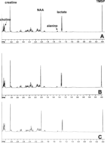
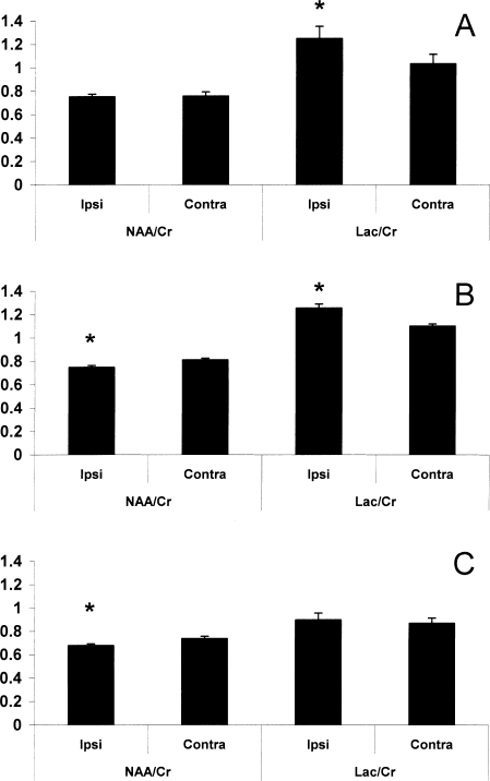
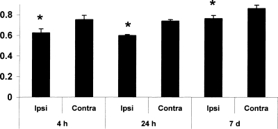
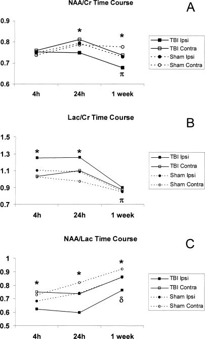
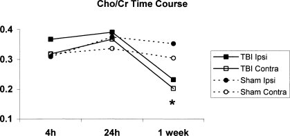
Although studies have shown alterations in cerebral metabolism after traumatic brain injury (TBI), clinical data in the developing brain is limited. We hypothesized that post-traumatic metabolic changes occur early (<24 h) and persist for up to 1 week. Immature rats underwent TBI to the left parietal cortex. Brains were removed at 4 h, 24 h, and 7 days after injury, and separated into ipsilateral (injured) and contralateral (control) hemispheres. Proton nuclear magnetic resonance (NMR) spectra were obtained, and spectra were analyzed for N-acetyl-aspartate (NAA), lactate (Lac), creatine (Cr), choline, and alanine, with metabolite ratios determined (NAA/Cr, Lac/Cr). There were no metabolic differences at any time in sham controls between cerebral hemispheres. At 4 and 24 h, there was an increase in Lac/Cr, reflecting increased glycolysis and/or decreased oxidative metabolism. At 24 h and 7 days, there was a decrease in NAA/Cr, indicating loss of neuronal integrity. The NAA/Lac ratio was decreased ( approximately 15-20%) at all times (4 h, 24 h, 7 days) in the injured hemisphere of TBI rats. In conclusion, metabolic derangements begin early (<24 h) after TBI in the immature rat and are sustained for up to 7 days. Evaluation of early metabolic alterations after TBI could identify novel targets for neuroprotection in the developing brain.
Figures
References
-
- Ashwal S. Holshouser B.A. Tomasi L.G. Shu S. Perkin R.M. Nystrom G.A. Hinshaw D.B., Jr. 1H-Magnetic resonance spectroscopy–determined cerebral lactate and poor neurological outcomes in children with central nervous system disease. Ann. Neurol. 1997;41:470–481. - PubMed
-
- Ashwal S. Holshouser B.A. Shu S.K. Simmons P.L. Perkin R.M. Tomasi L.G. Knierim D.S. Sheridan C. Craig K. Andrews G.H. Hinshaw D.B. Predictive value of proton magnetic resonance spectroscopy in pediatric closed head injury. Pediatr. Neurol. 2000;23:114–125. - PubMed
-
- Ashwal S. Holshouser B. Tong K. Serna T. Osterdock R. Gross M. Kido D. Proton MR spectroscopy detected glutamate/glutamine is increased in children with traumatic brain injury. J. Neurotrauma. 2004;21:1539–1552. - PubMed
-
- Ashwal S. Babikian T. Gardner-Nichols J. Freier M.C. Tong K.A. Holshouser B.A. Susceptibility-weighted imaging and proton magnetic resonance spectroscopy in assessment of outcome after pediatric traumatic brain injury. Arch. Phys. Med. Rehabil. 2006a;87:S50–S58. - PubMed
-
- Ashwal S. Holshouser B.A. Tong K.A. Use of advanced neuroimaging techniques in the evaluation of pediatric traumatic brain injury. Dev. Neurosci. 2006b;28:309–326. - PubMed
Publication types
MeSH terms
Substances
Grants and funding
LinkOut - more resources
Full Text Sources
Medical
