Review
. 2008 Sep-Oct;41(5):421-43.
doi: 10.1016/j.jcomdis.2008.03.004. Epub 2008 Mar 25.

Genetic and pharmacological intervention for treatment/prevention of hearing loss

Affiliations
Review

Genetic and pharmacological intervention for treatment/prevention of hearing loss

Douglas A Cotanche. J Commun Disord. 2008 Sep-Oct.

Abstract

Twenty years ago it was first demonstrated that birds could regenerate their cochlear hair cells following noise damage or aminoglycoside treatment. An understanding of how this structural and functional regeneration occurred might lead to the development of therapies for treatment of sensorineural hearing loss in humans. Recent experiments have demonstrated that noise exposure and aminoglycoside treatment lead to apoptosis of the hair cells. In birds, this programmed cell death induces the adjacent supporting cells to undergo regeneration to replace the lost hair cells. Although hair cells in the mammalian cochlea undergo apoptosis in response to noise damage and ototoxic drug treatment, the supporting cells do not possess the ability to undergo regeneration. However, current experiments on genetic manipulation, gene therapy, and stem cell transplantation suggest that regeneration in the mammalian cochlea may eventually be possible and may 1 day provide a therapeutic tool for hearing loss in humans.

Learning outcomes: The reader should be able to: (1) Describe the anatomy of the avian and mammalian cochlea, identify the individual cell types in the organ of Corti, and distinguish major features that participate in hearing function, (2) Demonstrate a knowledge of how sound damage and aminoglycoside poisoning induce apoptosis of hair cells in the cochlea, (3) Define how hair cell loss in the avian cochlea leads to regeneration of new hair cells and distinguish this from the mammalian cochlea where there is no regeneration following damage, and (4) Interpret the potential for new approaches, such as genetic manipulation, gene therapy and stem cell transplantation, could provide a therapeutic approach to hair cell loss in the mammalian cochlea.


Figures

Fig. 1
Fig. 1
The human cochlea. A classic anatomical drawing from Frank Netter of a mid-modiolar cross-section through the human cochlea showing the centrally located cochlear spiral ganglion and the various coils of the cochlear duct. The higher magnification drawing on the left represents a cross-section through an individual turn and shows the location of the organ of Corti, the three scalae and the peripheral process of the cochlear nerve cells. Used with permission from the Netter Atlas of Human Anatomy, 4th Edition.
Fig. 2
Fig. 2
The organ of corti. A confocal micrograph of the surface of the organ of Corti from a mouse cochlea showing the four rows of hair cells. This preparation was labeled with fluorescent phalloidin, a marker for actin filaments in cells. The intense labeling of the stereociliary bundle on each hair cell reveals the actin bundles that form the structural core of the stereocilia. IHC, inner hair cell; OHC1, first row of outer hair cells; OHC2, second row of outer hair cells; OHC3, third row of outer hair cells.
Fig. 3
Fig. 3
The avian cochlea. (A) This light micrograph shows an isolated preparation of the entire chick cochlea. The lateral stripes seen on the upper surface represent the folds of the tegmentum vasculosum, the ion-transporting epithelium in the chick cochlea. At the distal tip of the avian cochlea is the horseshoe-shaped lagena, a low-frequency sound and vibration detector. (B) This low power confocal micrograph shows a surface preparation of the basilar papilla, the sensory epithelium of the avian cochlea. The chick cochlea has a tonotopic organization, where high frequencies are heard in the narrow, proximal part to the left, and low frequencies are heard at the wide, distal end shown to the right. This preparation is labeled with fluorescent phalloidin, marking the actin filaments in the hair cells and supporting cells.
Fig. 4
Fig. 4
Gentamicin damage in the chick cochlea. These two low power confocal micrographs show only the high frequency half of the bird cochlea. A normal high frequency region from a control bird is shown in A. The white dots represent hair cells. A gentamicin treated bird cochlea is shown in B, at 72 h after a single injection of gentamicin. There are no remaining hair cells in the high frequency end of this cochlea, as seen by the loss of the while dots (arrows). Regenerating hair cells in this region are not yet identifiable.
Fig. 5
Fig. 5
Apoptosis in hair cells. This low power confocal micrograph shows the proximal, high frequency region of the chick cochlea 72 h after gentamicin treatment. The dying hair cells contain activated caspase-3 (green), the executioner caspase in apoptosis. Surviving hair cells and other cell types in the cochlea are labeled with phalloidin (red) to detect actin filaments.
Fig. 6
Fig. 6
Apoptotic proteins in the death cascade. The earliest sign that apoptosis has been activated following gentamicin treatment is the translocation of TIAR out of the nucleus into the cytoplasm of hair cells. A normal cochlea is shown in A, where the TIAR is localized to all the cell nuclei of both hair cells and supporting cells (inset). In B, the TIAR in hair cells move out of the nucleus and forms brightly fluorescent granules in the hair cell cytoplasm (inset) only 24 h after gentamicin treatment. The next identifiable step in apoptosis is the release of cytochrome c from mitochondria into the hair cell cytoplasm. In C the distribution of cytochrome c is shown (green) in hair cells double-labeled with phalloidin (red). In D only the distribution of cytochrome c is shown for the same sample. The normal, punctate distribution of cytochrome c in mitochondria is seen in the two cells at the very top of the micrograph. In the cells labeled with arrows cytochrome c has spilled out into the hair cell cytoplasm and fills it with a green fluorescence. Finally the executioner stage of apoptosis is reached with the activation of caspase-3. In E and F the distribution of the activated form of caspase 3 is shown in hair cells that are in the process of blebbing up and being ejected from the cochlear epithelium (at arrows, plus in other cells). The green label in E and F is the activated form of caspase 3. The red label in E represents phalloidin labeling of the hair cells.
Fig. 7
Fig. 7
Hair cell regeneration in the chick cochlea. This whole-mount confocal micrograph shows the development of new hair cells in the chick cochlea 7 days after a single gentamicin treatment. The new hair cells (green) are labeled with a marker for myosin VI, a protein found only in hair cells in the cochlear sensory epithelium. Some of the new hair cells are labeled with BrdU, a marker for DNA synthesis in dividing cells. The new hair cells that lack BrdU in their nuclei were generated by direct transdifferentiation (white arrows), while those that have BrdU-labeled nuclei were made through the mitosis of a supporting cell. Some labeled nuclei are in newly generated supporting cells, which don't label with myosin VI (red arrows).
Fig. 8
Fig. 8
Stem cell transplantation into the cochlear nerve. Male mouse neural stem cells were transplanted into female guinea pigs allowed to integrate into the cochlear nerve bundle for 6 weeks. In A, labeling with a marker for the male Y chromosome (green dot) showed that both satellite cells (white arrows) and neurons (green arrow) in the recovered cochlear nerve were derived from stem cells. Neurons are labeled with neurofilament 200 (red), a protein found only in neurons. All cell nuclei are labeled with DAPI (light blue), a marker for DNA. In B, a satellite cell derived from a stem cell (yellow arrow) is labeled with S100b (red), a protein found satellite cells and not neurons. In C, a neuron in the cochlear spiral ganglion derived from a stem cell (green arrow) is labeled with neurofilament 200 (red). Cell nuclei in B and C are labeled with DAPI.
Fig. 9
Fig. 9
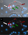
Stem cell transplantation into the organ of corti. The two micrographs in A and B represent a brightfield/ fluorescence view (A) and a fluorescence-only view of the same section through a cochlea that was transplanted with stem cells. The third row outer hair cell in these images is labeled with a male Y chromosome marker (white arrow), while the first and second row outer hair cells (green arrow) and the inner hair cell (red arrow) are not. The hair cells are also labeled for Myosin VIIa, a marker found only in hair cells in the cochlea. All cell nuclei are labeled with DAPI (light blue).

References

    1. Adams JM, Cory S. The Bcl-2 protein family: Arbiters of cell survival. Science. 1998;281:1322–1326. - PubMed
    1. Bermingham NA, Hassan BA, Price SD, Vollrath MA, Ben-Arie N, Eatock RA, et al. Math1: An essential gene for the generation of inner ear hair cells. Science. 1999;284:1837–1841. - PubMed
    1. Bhave SA, Stone JS, Rubel EW, Coltrera MD. Cell cycle progression in gentamicin-damaged avian cochleas. Journal of Neuroscience. 1995;15:4618–4628. - PMC - PubMed
    1. Cafaro J, Lee GS, Stone JS. Atoh1 expression defines activated progenitors and differentiating hair cells during avian hair cell regeneration. Developmental Dynamics. 2007;236:156–170. - PubMed
    1. Campbell K, Rybak L. Otoprotective agents. In: Campbell KCM, editor. Pharmacology and ototoxicity for audiologists. Thomson Delmar Learning; Clifton Park, NY: 2007. pp. 287–300.

Publication types

MeSH terms

Substances