

Unraveling the microenvironmental influences on the normal mammary gland and breast cancer
- PMID: 18455428
- PMCID: PMC2548304
- DOI: 10.1016/j.semcancer.2008.03.013
Unraveling the microenvironmental influences on the normal mammary gland and breast cancer
Abstract
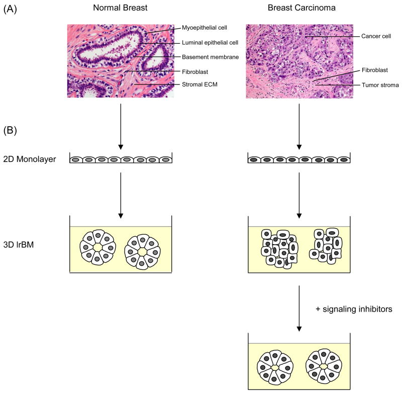
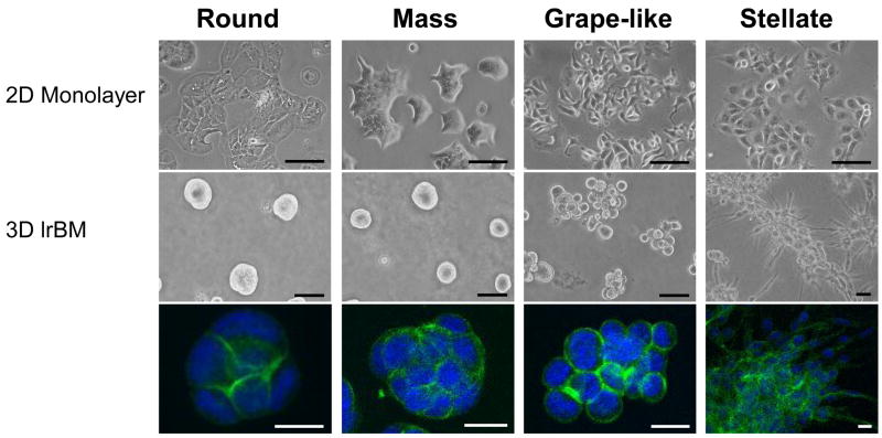
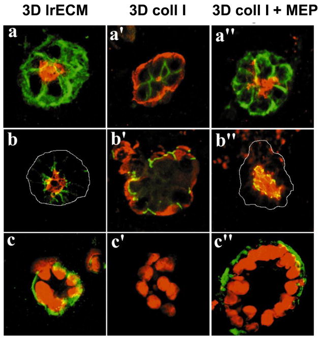
The normal mammary gland and invasive breast tumors are both complex 'organs' composed of multiple cell types as well as extracellular matrix in three-dimensional (3D) space. Conventionally, both normal and malignant breast cells are studied in vitro as two-dimensional monolayers of epithelial cells, which results in the loss of structure and tissue function. Many laboratories are now investigating regulation of signaling function in the normal mammary gland using 3D cultures. However, it is also important to assay malignant breast cells ex vivo in a physiologically relevant environment to more closely mimic tumor architecture, signal transduction regulation and tumor behavior in vivo. Here we present the potential of these 3D models for drug testing, target validation and guidance of patient selection for clinical trials. We also argue that in order to get full insight into the biology of the normal and malignant breast, and to create in vivo-like models for therapeutic approaches in humans, we need to continue to create more complex heterotypic models to approach the full context the cells encounter in the human body.
Figures
Similar articles
-
Mammary development and breast cancer: the role of stem cells.Curr Mol Med. 2011 Jun;11(4):270-85. doi: 10.2174/156652411795678007. Curr Mol Med. 2011. PMID: 21506923 Free PMC article. Review.
-
Stem cells in normal mammary gland and breast cancer.Am J Med Sci. 2010 Apr;339(4):366-70. doi: 10.1097/MAJ.0b013e3181cad964. Am J Med Sci. 2010. PMID: 20224316 Review.
-
Normal and cancerous mammary stem cells evade interferon-induced constraint through the miR-199a-LCOR axis.Nat Cell Biol. 2017 Jun;19(6):711-723. doi: 10.1038/ncb3533. Epub 2017 May 22. Nat Cell Biol. 2017. PMID: 28530657 Free PMC article.
-
Wnt signaling, stem cells, and the cellular origin of breast cancer.Stem Cell Rev. 2007 Jun;3(2):157-68. doi: 10.1007/s12015-007-0025-3. Stem Cell Rev. 2007. PMID: 17873348 Review.
-
Harnessing 3D models of mammary epithelial morphogenesis: An off the beaten path approach to identify candidate biomarkers of early stage breast cancer.Cancer Lett. 2016 Oct 1;380(2):375-383. doi: 10.1016/j.canlet.2016.07.003. Epub 2016 Jul 12. Cancer Lett. 2016. PMID: 27422542 Free PMC article.
Cited by
-
Spheroid Model of Mammary Tumor Cells: Epithelial-Mesenchymal Transition and Doxorubicin Response.Biology (Basel). 2024 Jun 21;13(7):463. doi: 10.3390/biology13070463. Biology (Basel). 2024. PMID: 39056658 Free PMC article.
-
Production of Uniform 3D Microtumors in Hydrogel Microwell Arrays for Measurement of Viability, Morphology, and Signaling Pathway Activation.Assay Drug Dev Technol. 2015 Nov;13(9):570-83. doi: 10.1089/adt.2015.662. Epub 2015 Aug 14. Assay Drug Dev Technol. 2015. PMID: 26274587 Free PMC article.
-
Organotypic 3D cell culture models: using the rotating wall vessel to study host-pathogen interactions.Nat Rev Microbiol. 2010 Nov;8(11):791-801. doi: 10.1038/nrmicro2423. Nat Rev Microbiol. 2010. PMID: 20948552 Review.
-
Tumor cells and their crosstalk with endothelial cells in 3D spheroids.Sci Rep. 2017 Sep 5;7(1):10428. doi: 10.1038/s41598-017-10699-y. Sci Rep. 2017. PMID: 28874803 Free PMC article.
-
Bone: A Fertile Soil for Cancer Metastasis.Curr Drug Targets. 2017;18(11):1281-1295. doi: 10.2174/1389450117666161226121650. Curr Drug Targets. 2017. PMID: 28025941 Free PMC article. Review.
References
Publication types
MeSH terms
Substances
Grants and funding
LinkOut - more resources
Full Text Sources
Other Literature Sources
Medical
