. 2008 Jun 15;318(2):276-88.
doi: 10.1016/j.ydbio.2008.03.028. Epub 2008 Mar 28.

Fibroblast growth factor receptor signaling is essential for lens fiber cell differentiation

Affiliations

Fibroblast growth factor receptor signaling is essential for lens fiber cell differentiation

Haotian Zhao et al. Dev Biol. .

Abstract

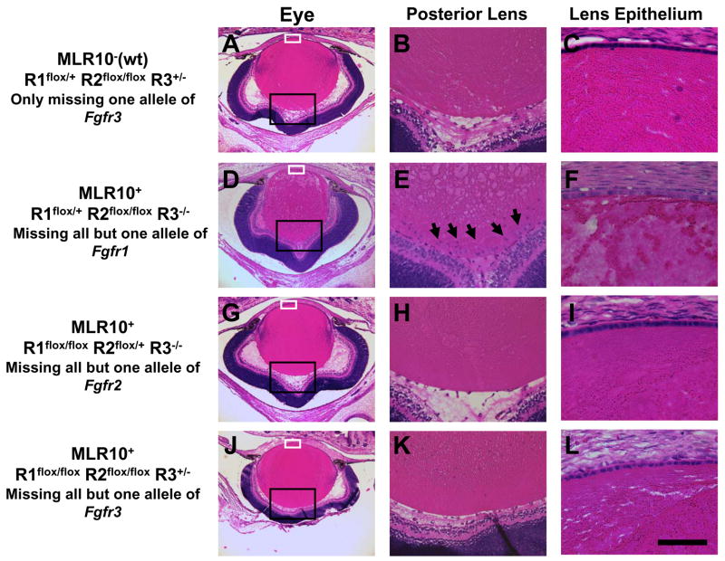
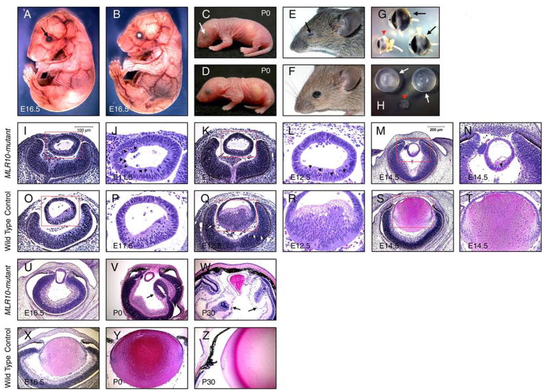
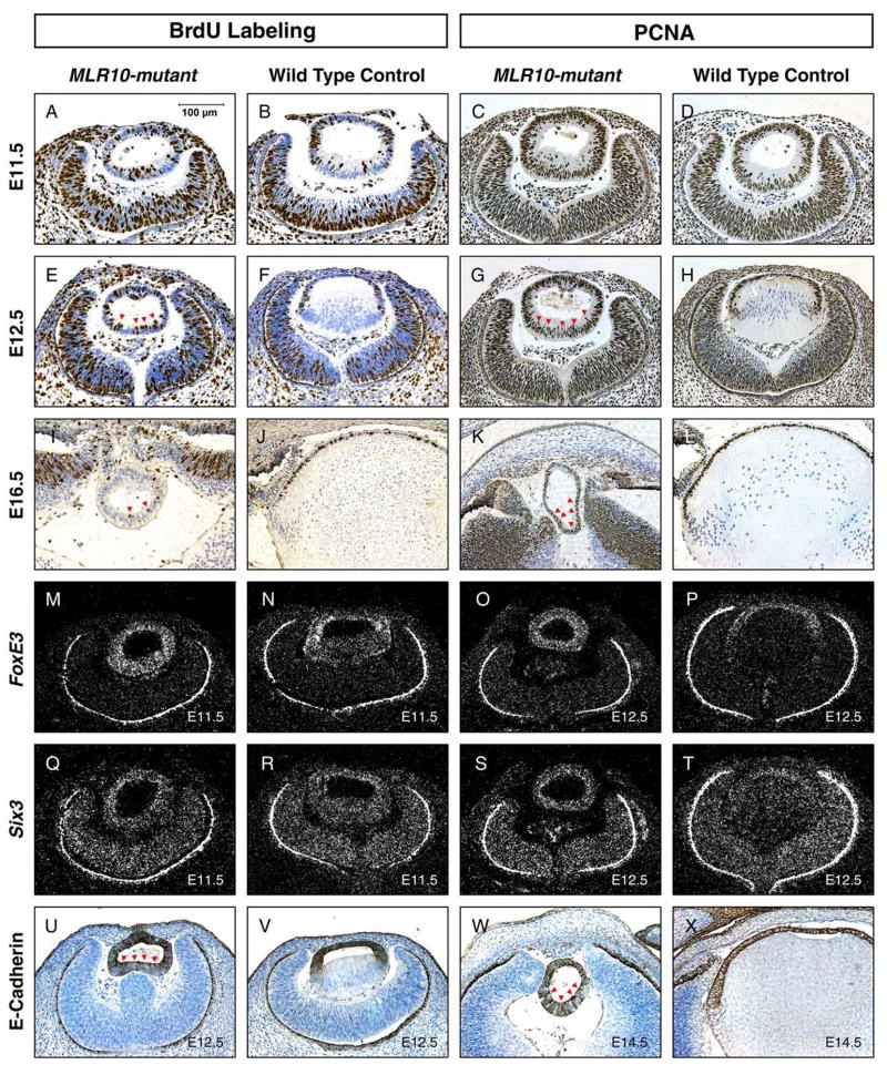
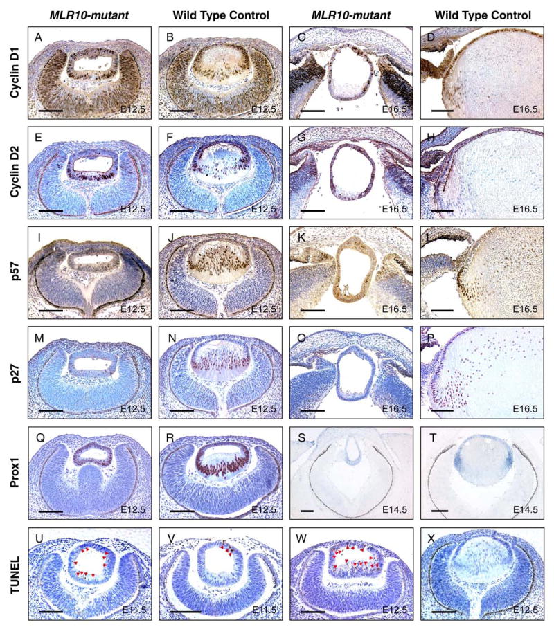
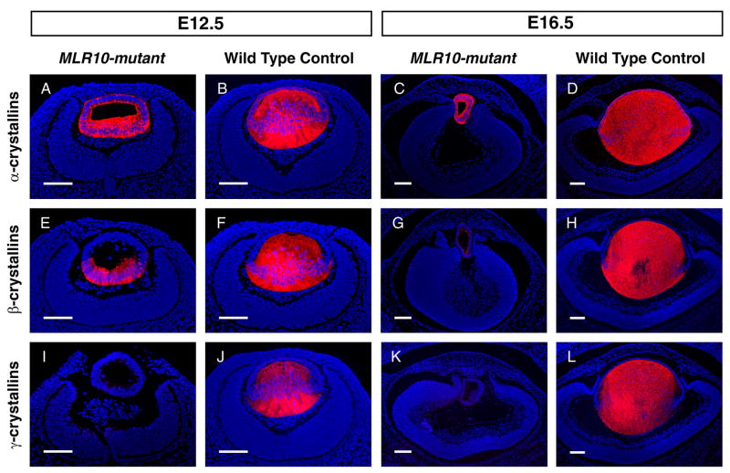
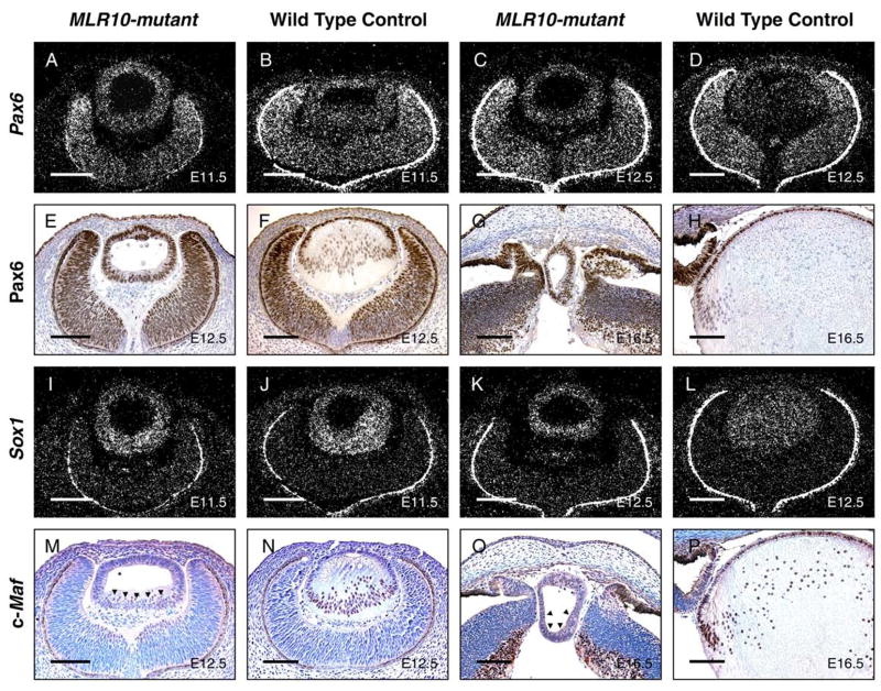
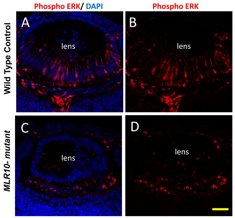
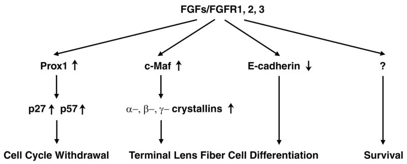
The vertebrate lens provides an excellent model to study the mechanisms that regulate terminal differentiation. Although fibroblast growth factors (FGFs) are thought to be important for lens cell differentiation, it is unclear which FGF receptors mediate these processes during different stages of lens development. Deletion of three FGF receptors (Fgfr1-3) early in lens development demonstrated that expression of only a single allele of Fgfr2 or Fgfr3 was sufficient for grossly normal lens development, while mice possessing only a single Fgfr1 allele developed cataracts and microphthalmia. Profound defects were observed in lenses lacking all three Fgfrs. These included lack of fiber cell elongation, abnormal proliferation in prospective lens fiber cells, reduced expression of the cell cycle inhibitors p27(kip1) and p57(kip2), increased apoptosis and aberrant or reduced expression of Prox1, Pax6, c-Maf, E-cadherin and alpha-, beta- and gamma-crystallins. Therefore, while signaling by FGF receptors is essential for lens fiber differentiation, different FGF receptors function redundantly.


Figures

Figure 1
Figure 1
Lens development when five of the six Fgfr1-3 alleles are missing. Sections from newborn eyes from animals missing just one allele of Fgfr3 (A–C) are compared with those missing all FGF receptors, except one allele of Fgfr1 (D–F), all except one allele of Fgfr2 (G–I) or all except one allele of Fgfr3 (J–L). Regions boxed in black and white in the first column (A, D, G, J) are shown at higher magnification in the second (B, E, H, K) and third (C, F, I, L) columns, respectively. Notice that there is an accumulation of nucleated cells at the posterior region of the lens containing only one allele of Fgfr1 (arrows, E). This genotype is also typified by fiber cell degeneration and a lower than normal density of lens epithelial cells (F). MLR10-designates mice in which the MLR10 transgene was not present. R1, R2 and R3 represent Fgfr1, Fgfr2 and Fgfr3 respectively. The conditional, null and wild type (wt) alleles of these genes are represented by flox, −, and + respectively. The scale bar in (L) represents 50 μm in (C, F, I, L), 125 μm in (B, E, H, K) and 500 μm in (A, D, G, J).
Figure 2
Figure 2
Defective lens fiber elongation in MLR10-mutant (MLR10/Fgfr1flox/flox Fgfr2flox/flox Fgfr3 /) mice. A–F: MLR10-mutant mice (A, C, E) were compared with control mice (B, D, F) at E16.5 (A, B), P0 (C, D) and P30 (E, F). Triple Fgfr-deficient mice were characterized by severe microphthalmia (arrows A, C, E); G: Eyes of MLR10-mutant (arrowhead, G) were placed together with control eyes (arrows, G). The anterior chamber (*) is absent in the mutant eye; H: Lenses from MLR10-mutant mice (arrowhead, H) were much smaller than control lenses (arrows, H); I-Z: Histological analysis of MLR10-mutant (I–N, U–W) and control (O–T, X–Z) eyes. Developmental stages studied include E11.5 (I, J, O, P), E12.5 (K, L, Q, R), E14.5 (M, N, S, T) E16.5 (U, X), P0 (V, Y) and P30 (W, Z). The boxed regions in I, O, K, Q, M, S are shown at higher magnification in J, P, L, R, N, T respectively and pyknotic nuclei are indicated by arrowheads (J, L, N). Arrows in V and W indicate abnormal folds of neural retina in the mutant eyes.
Figure 3
Figure 3
Defects in cell cycle exit and persistent expression of lens epithelial markers in MLR10-mutant lenses. A-L: BrdU-incorporation (A, B, E, F, I, J) and PCNA expression (C, D, G, H, K, L) analyses were performed on mutant (A, E, I, C, G, K) and control (B, F, J, D, H, L) embryos at E11.5 (A–D), E12.5 (E–H) and E16.5 (I–L). Brown nuclear staining indicated cells that were in the S-phase of cell cycle (1st and 2nd column, A–L), during BrdU labeling, or that express PCNA (3rd and 4th column, A-L), typical of proliferating cells. Red arrowheads mark BrdU-incorporating or PCNA-expressing mutant cells in the posterior portion of lenses. M–T: In-situ hybridization analyses of lens epithelial markers FoxE3 (M–P), Six3 (Q–T) at E11.5 (M, N, Q, R) and E12.5 (O, P, S, T) were performed on mutant (M, Q, O, S) and control (N, R, P, T) lenses. Bright-appearing silver grains in the dark field photos indicate expression of these genes. The retinal pigmented epithelium (RPE) appears as a bright line surrounding the optic cup in the darkfield illumination, due to light scattering by pigment granules. U–X: Both mutant (U, W) and control (V, X) lenses at E12.5 day (U, V) and E14.5 day (W, X) were analyzed for the expression of the lens epithelial marker E-cadherin by immunohistochemistry. Dark brown staining indicates E-cadherin expression in junctions between lens epithelial cells. Red arrowheads mark cells expressing E-cadherin in the posterior of the MLR10-mutant lens.
Figure 4
Figure 4
Analysis of the expression of cell cycle regulators and cell death in MLR10-mutant lenses. Cyclin D1 (A–D), cyclin D2 (E–H), p57kip2 (I-L), p27kip1 (M–P) and Prox1 (Q–R) levels were analyzed by immunohistochemistry in the lenses of MLR10-mutant (A, E, I, M, C, G, K, O, Q) and control (B, F, J, N, D, H, L, P, R) mice. Prox1 mRNA expression was also examined by in situ hybridization in MLR10-mutant and control lenses (S and T, respectively). TUNEL assays were conducted on mutant (U, W) and control (V, X) embryonic lenses. Developmental stages studied included E11.5 (U,V) E12.5 (A, B, E, F, I, J, M, N, Q, R, W, X), E14.5 (S, T) and E16.5 (C, D, G, H, K, I, O, P). Brown nuclear staining in A–D, I–L, U–W, purplish nuclear staining in E–H and M–R and dark blue staining in S and T indicated positive staining for the relevant protein or mRNA. Arrowheads in U, V, W marked apoptotic cells in MLR10-mutant and control lenses. Note that TUNEL-positive cells were detected throughout the mutant lens. All scale bars = 100 μm.
Figure 5
Figure 5
Impaired crystallin expression in MLR10–mutant lenses. Immunofluorescence analyses of α-crystallins (A–D), β-crystallins (E–H) and γ-crystallins (I–L) were carried out in MLR10–mutant (A, C, E, G, I, K) and control (B, D, F, H, J, L) animals. Developmental stages studied included E12.5 (A, B, E, F, I, J) and E16.5 (C, D, G, H, K, L). Red-fluorescence indicates positive antibody staining. DAPI stained nuclei are blue. All scale bars = 100 μm.
Figure 6
Figure 6
Analysis of the expression of transcription factors required for crystallin expression in MLR10–mutant lenses. In-situ hybridization (A–D, I–L) and immunohistochemistry (E–H, M–P) revealed the expression of Pax6 (A–H), Sox1 (I–L) and c-Maf (M-P) in mutant (A, C, E, G, I, K, M, O) and control (B, D, F, H, J, L, N, P) lenses. Developmental stages studied included E11.5 (A, B, I, J), E12.5 (C, D, E, F K, L, M, N) and E16.5 (G, H, O, P). For pictures of immunohistochemistry, brown nuclear staining in E–H and purplish nuclear staining in M–P indicate positive staining. Bright silver grains reveal hybridization signals from relevant transcripts. The retinal pigmented epithelium (RPE) appears as a bright line surrounding the optic cup, due to light scattering by pigment granules. Note that the Pax6 staining in the nuclei of both the anterior and posterior cells of the MLR10-mutant lenses (E and G) while Pax6 staining in the wild type lens is largely restricted to anterior lens epithelial cells (F and H). Black arrowheads in M and O represent sparse nuclei expressing c-Maf in the MLR10-mutant lens. All scale bars = 100 μm.
Figure 7
Figure 7
Deletion of Fgfrs leads to reduction in phosphorylated Erk1/2 in the lens. Phosphorylated (active) forms of Erk1 and Erk2 were not evident in lens epithelial cells, but were readily detected in elongating primary fiber cells in control lenses (A, B). Phospho-Erk1/2 staining was dramatically reduced in the MLR10-mutant lenses (C and D) at E12.5. Nuclei were counterstained with DAPI (blue), with phosphorylated Erk1/2 staining appearing red. Scale bar = 50 μm.
Figure 8
Figure 8
A model for the coordination of cell cycle withdrawal and lens fiber differentiation by FGF signaling. Prox1 is required for the increased expression of p27kip1 and p57kip2 that mediates the entry of lens fiber cells into G0. Deficient FGF receptor signaling led to reduced expression of Prox1 and decreased expression of p27kip1 and p57kip2, resulting in the abnormal proliferation of cells that would normally form lens fibers. The transcription factor c-Maf promotes the expression of α-, β- and γ-crystallins and fiber cell elongation. FGF signaling deficiency led to decreased crystallin gene expression and failure of fiber cell elongation. FGF signaling is also required for the decrease in E-cadherin expression that normally accompanies fiber cell differentiation. Finally, increased apoptosis in the MLR10-mutant lenses suggests that FGF signaling is required for lens epithelial cell survival.

Similar articles

Cited by

References

    1. Arman E, Haffner-Krausz R, Chen Y, Heath JK, Lonai P. Targeted disruption of fibroblast growth factor (FGF) receptor 2 suggests a role for FGF signaling in pregastrulation mammalian development. Proc Natl Acad Sci U S A. 1998;95:5082–7. - PMC - PubMed
    1. Ashery-Padan R, Marquardt T, Zhou X, Gruss P. Pax6 activity in the lens primordium is required for lens formation and for correct placement of a single retina in the eye. Genes Dev. 2000;14:2701–11. - PMC - PubMed
    1. Belecky-Adams TL, Adler R, Beebe DC. Bone morphogenetic protein signaling and the initiation of lens fiber cell differentiation. Development. 2002;129:3795–802. - PubMed
    1. Blixt A, Mahlapuu M, Aitola M, Pelto-Huikko M, Enerback S, Carlsson P. A forkhead gene, FoxE3, is essential for lens epithelial proliferation and closure of the lens vesicle. Genes Dev. 2000;14:245–54. - PMC - PubMed
    1. Chow RL, Roux GD, Roghani M, Palmer MA, Rifkin DB, Moscatelli DA, Lang RA. FGF suppresses apoptosis and induces differentiation of fibre cells in the mouse lens. Development. 1995;121:4383–4393. - PubMed

Publication types

MeSH terms

Substances