. 2008 Jun 6;379(3):505-19.
doi: 10.1016/j.jmb.2008.03.075. Epub 2008 Apr 8.

Mutations outside the anisomycin-binding site can make ribosomes drug-resistant

Affiliations

Mutations outside the anisomycin-binding site can make ribosomes drug-resistant

Gregor Blaha et al. J Mol Biol. .

Abstract

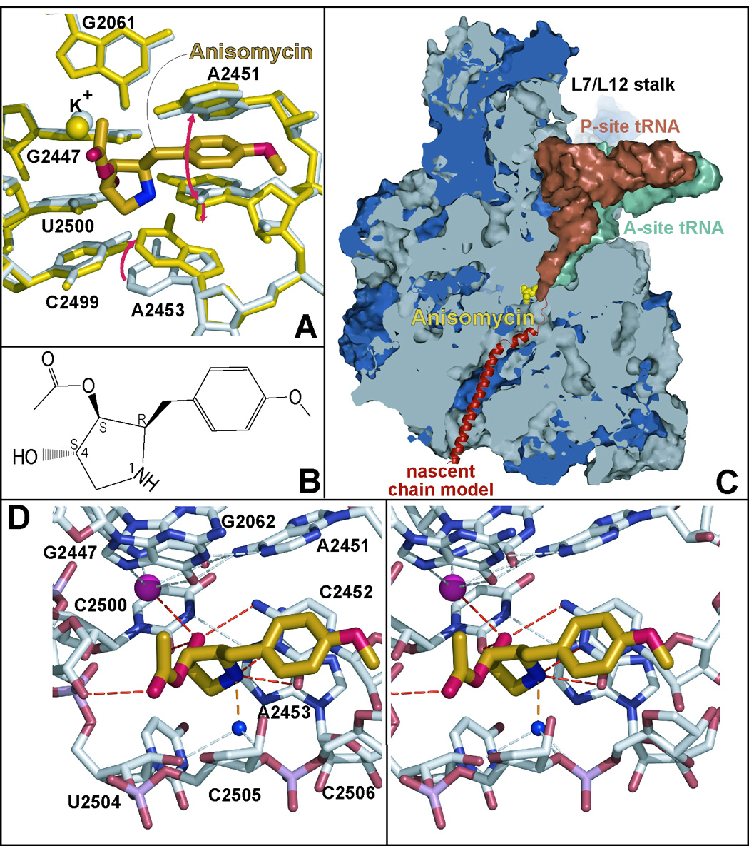
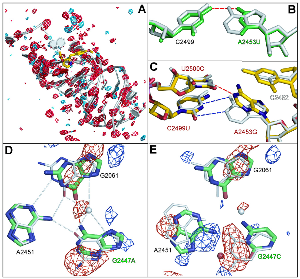
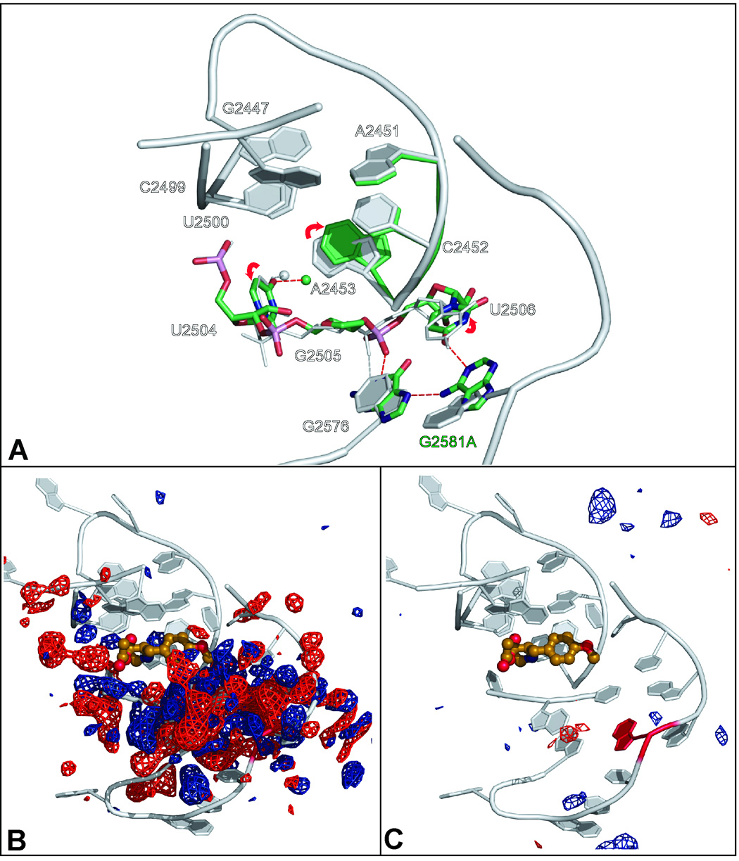
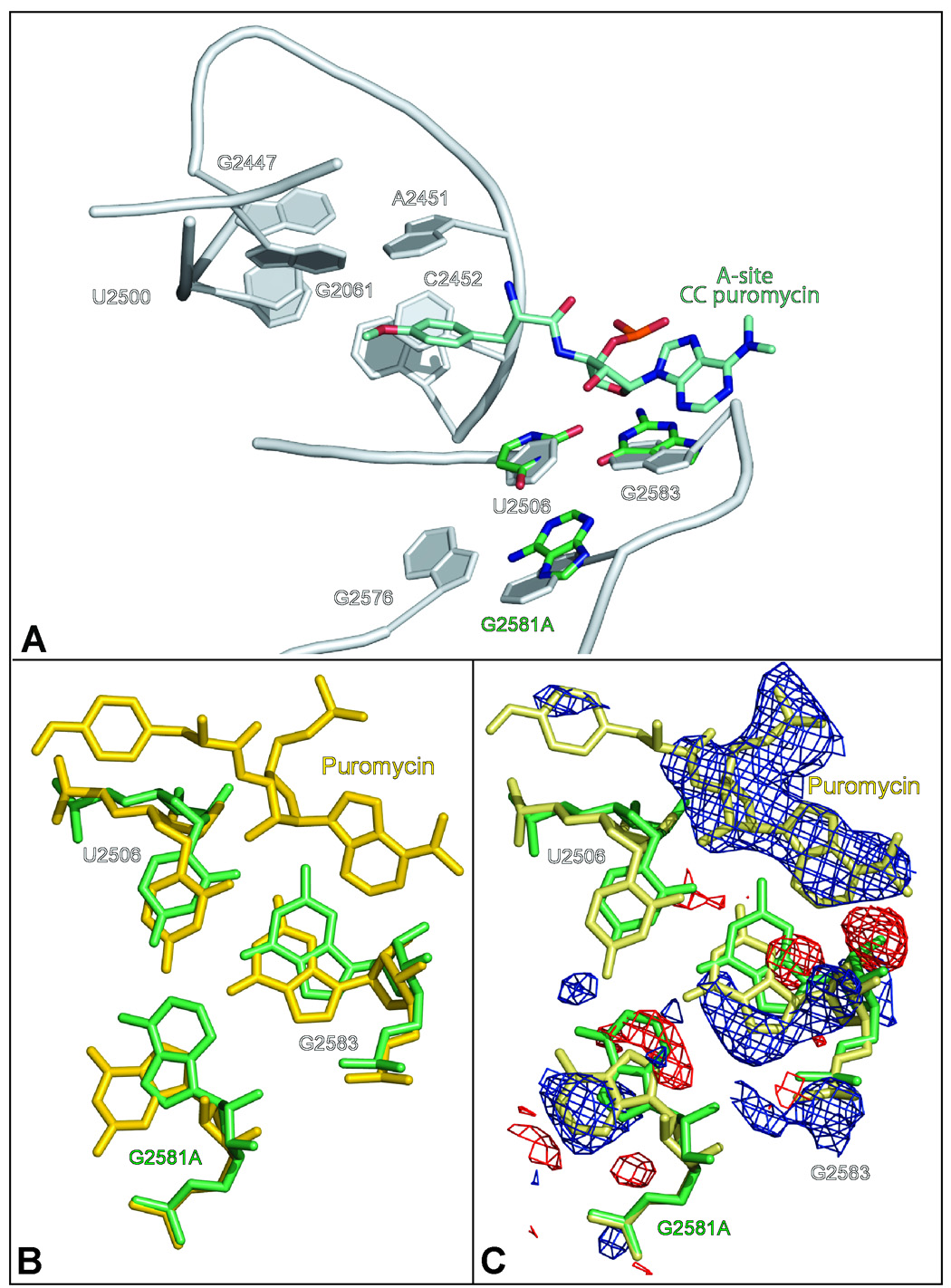
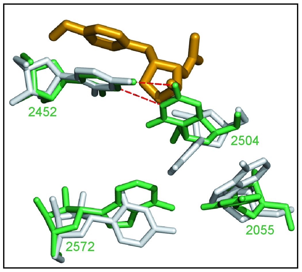
Eleven mutations that make Haloarcula marismortui resistant to anisomycin, an antibiotic that competes with the amino acid side chains of aminoacyl tRNAs for binding to the A-site cleft of the large ribosomal unit, have been identified in 23S rRNA. The correlation observed between the sensitivity of H. marismortui to anisomycin and the affinity of its large ribosomal subunits for the drug indicates that its response to anisomycin is determined primarily by the binding of the drug to its large ribosomal subunit. The structures of large ribosomal subunits containing resistance mutations show that these mutations can be divided into two classes: (1) those that interfere with specific drug-ribosome interactions and (2) those that stabilize the apo conformation of the A-site cleft of the ribosome relative to its drug-bound conformation. The conformational effects of some mutations of the second kind propagate through the ribosome for considerable distances and are reversed when A-site substrates bind to the ribosome.


Figures

Figure 1
Figure 1
The location in the polypeptide exit tunnel of the large subunit where anisomycin binds to the ribosome and its interactions with its binding site. Figure 1A shows anisomycin (heavy gold structure with its elements in different colors) bound to the ribosome. The conformation of the nucleotides in its binding site when the drug is bound is shown in yellow. Their conformation in the absence of the drug is shown in gray. The most conspicuous base motions associated with drug binding are indicated by red arrows. Bases are numbered so that they correspond with the base numbering of the 23S rRNA of E. coli. In this case, as in all others shown here, structures are superimposed by aligning them on the positions of 50 phosphorus atoms in the peptidyl transferase center. Figure 1B presents the chemical structure of anisomycin. Figure 1C is a cut-away view of the large ribosomal subunit – rRNA’s in gray and the ribosomal proteins in blue – showing the A-site (green) and P-site (dull red) bound tRNAs, and a schematic representation of a nascent polypeptide in the peptide exit tunnel (red). The position occupied by anisomycin (gold) when bound to the ribosome is also shown. Figure 1D is a stereo pair showing the way anisomycin (gold) interacts with its binding site. The dashed lines indicate a potential hydrogen bond (orange), hydrogen bonds to anisomycin (red), and some hydrogen bonds detected within the large ribosomal subunit structure (gray).
Figure 2
Figure 2
A global view of the positions of the bases the mutation of which cause anisomycin-resistance in H. marismortui. The drug (gold with spherical atoms) is shown surrounded by the bases, the mutation of which lead to drug resistance (red). The backbone connecting bases is indicated in gray. The positions occupied by the CCA end of P-site bound tRNA (orange) and A-site bound tRNA (green) are shown for orientation. E. coli numbering is used for all bases.
Figure 3
Figure 3
The effects of mutations on the structure of the A-site cleft region. Figure 3A shows the difference electron density observed when wild type apo- data are subtracted from data obtained from crystals containing U2500A ribosomes. The contouring is at + 4 sigma (blue) and − 4 sigma (red). The correlation of red features with backbone phosphate groups is indicative of a general disordering of the structure in the neighborhood of the mutation. Figure 3B displays the effect of mutating A2453 (gray structure) to a U (green structure). Figure 3C overlays the wild type apo- structure (gray) with the wild type drug-bound structure (yellow) in the region of A2453, which is immediately below the A-site cleft. The mutations obtained in this region are indicated with red labels. Figure 3D shows how the mutation G2447A disturbs the structure of the (G2061, G2447, A2451) base triple. The mutant structure is in green and the wild type structure is in gray. The (FG2447A − Fwild type) electron difference map is contoured at a level of 6 sigma. The negative electron difference density is displayed in red and the positive density is in blue. Figure 3E is the corresponding figure for the mutation G2447C with the (FG2447C − Fwild type) electron difference density contoured at 4 sigma.
Figure 4
Figure 4
The effects of the mutation G2581A on the structure of the large ribosomal subunit from H. marismortui. Figure 4A compares the structure of the G2581A mutant (green) with the wild type apo structure (gray). Hydrogen bonds characteristic of the mutant are indicated by red dashed lines while hydrogen bond characteristic of the wild type structure of shown as gray dashed lines. Figure 4B shows the (FG2581A − Fwild type) electron density difference map computed from the amplitudes observed in G2581A crystals and the amplitudes obtained from wild type crystals. Contours are drawn at +4 sigma (blue) and − 4 sigma (red). The underlying structure is shown in gray. Figure 4C corresponds to Figure 4B, but the difference electron density map (FG2581A − FG2581A calculated) difference electron map shown was obtained using the observed amplitudes obtained from G2581A crystals and calculated amplitudes obtained from the refined structure for the mutated ribosome. Differences are contoured at the same levels as in Figure 4B.
Figure 5
Figure 5
The effect of A-site substrate binding on the conformation of large ribosomal subunits containing the mutation G2581A. Figure 5A compares the conformation of the 2581 region of wild type ribosomes (gray) with that of the G2581A mutant (green). Also included in the figure is CC-puromycin (bluegreen) as reference for the binding site of amino acylated tRNA to the A-site. Figure 5B compares the structure of G2581A mutant (green) and CC-puromycin bound to large ribosomal subunit of wild type (gold). Figure 5C compares the structures of G2581A mutant (green) with CC-puromycin bound to G2581A (khaki) with overlaid (FG2581A − FG2581A CC-puromycin) difference electron density, which was computed by using as amplitudes the differences observed between the data obtained from G2581A crystals that included the analog and data obtained from G2581A crystals that lack the analog. Positive features were contoured at +4 sigma (blue), and negative features at − 4 sigma (red).
Figure 6
Figure 6
A hypothesis for the structural difference that controls the response of different species to A-site cleft antibiotics. The structure of the T. thermophilus large ribosomal subunit in the neighborhood of C2504 (green) is compared to the structure of that same region in H. marismortui (gray) with anisomycin bound (gold).

References

    1. Nissen P, Hansen J, Ban N, Moore PB, Steitz TA. The structural basis of ribosome activity in peptide bond synthesis. Science. 2000;289:920–930. - PubMed
    1. Hansen JL, Schmeing TM, Moore PB, Steitz TA. Structural insights into peptide bond formation. Proc. Natl. Acad. Sci. USA. 2002;99:11670–11675. - PMC - PubMed
    1. Schmeing TM, Seila AC, Hansen JL, Freeborn B, Soukup JK, Scaringe SA, Strobel SA, Moore PB, Steitz TA. A pre-translocational intermediate in protein synthesis observed in crystals of enzymatically active 50S subunits. Nat. Struct. Biol. 2002;9:225–230. - PubMed
    1. Hansen JL, Moore PB, Steitz TA. Structures of five antibiotics bound at the peptidyl transferase center of the large ribosomal subunit. J. Mol. Biol. 2003;330:1061–1075. - PubMed
    1. Schlunzen F, Zarivach R, Harms J, Bashan A, Tocilj A, Albrecht R, Yonath A, Franceschi F. Structural basis for the interaction of antibiotics with the peptidyl transferase centre in eubacteria. Nature. 2001;413:814–821. - PubMed

Publication types

MeSH terms

LinkOut - more resources