

Transient elastic support for vein grafts using a constricting microfibrillar polymer wrap
- PMID: 18455787
- PMCID: PMC2486447
- DOI: 10.1016/j.biomaterials.2008.04.009
Transient elastic support for vein grafts using a constricting microfibrillar polymer wrap
Abstract
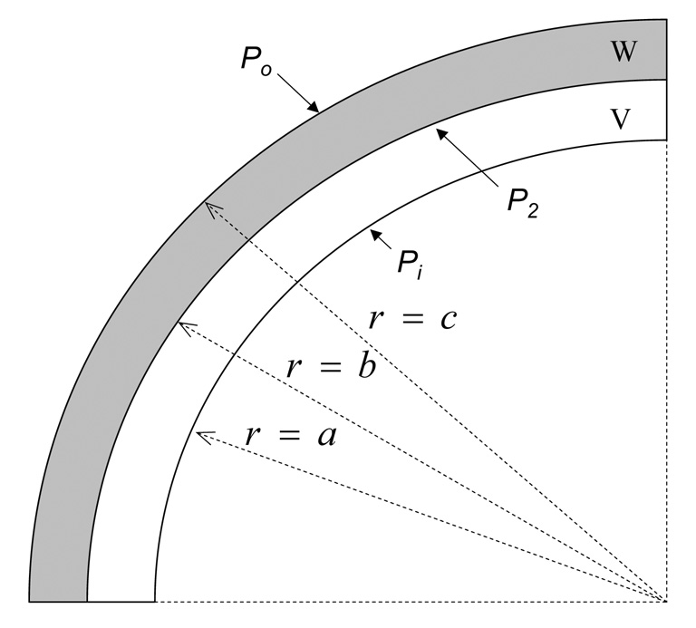
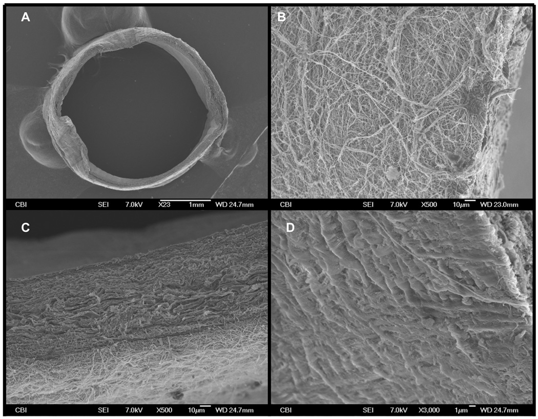
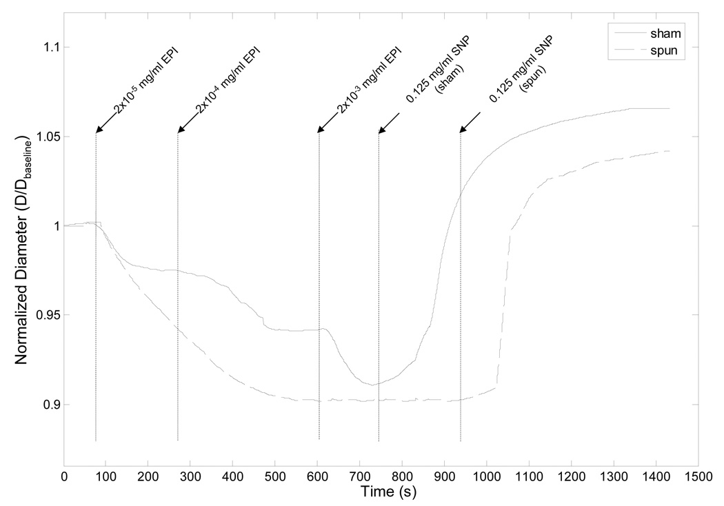
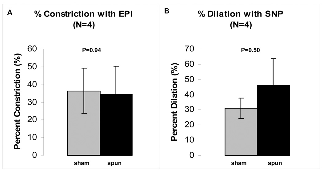
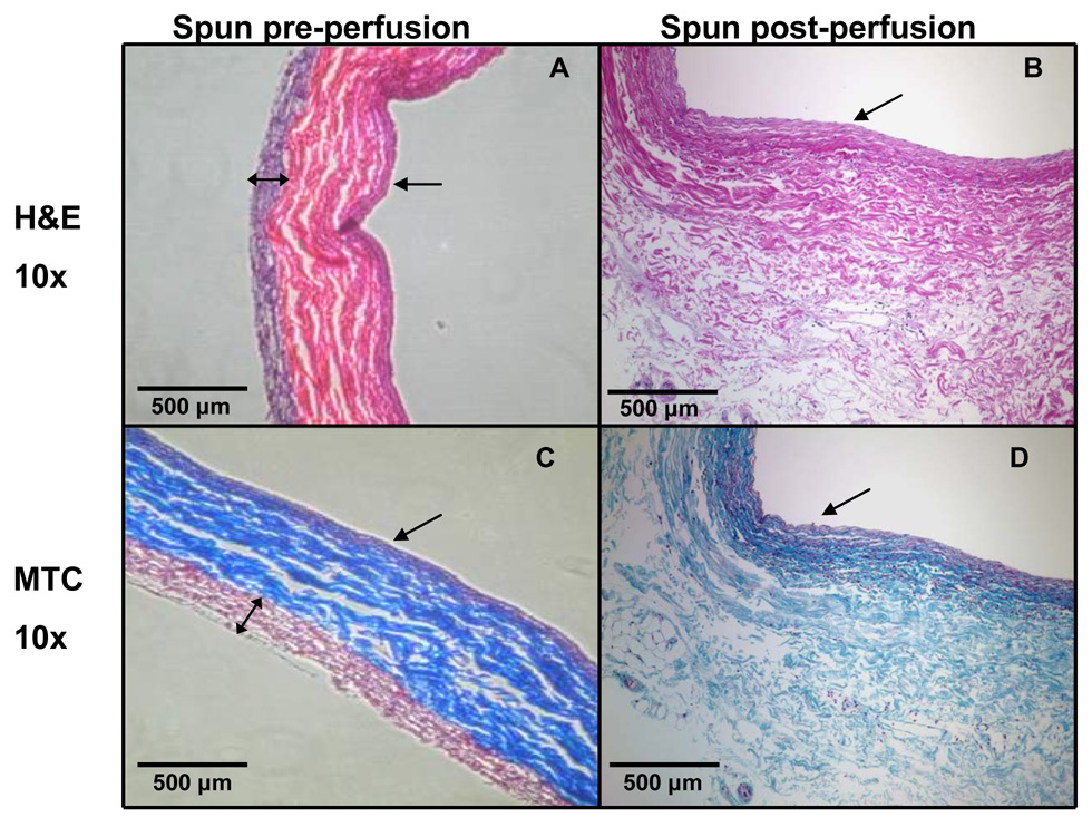
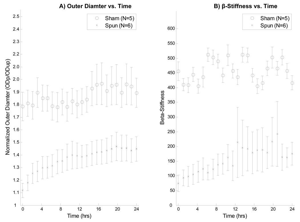
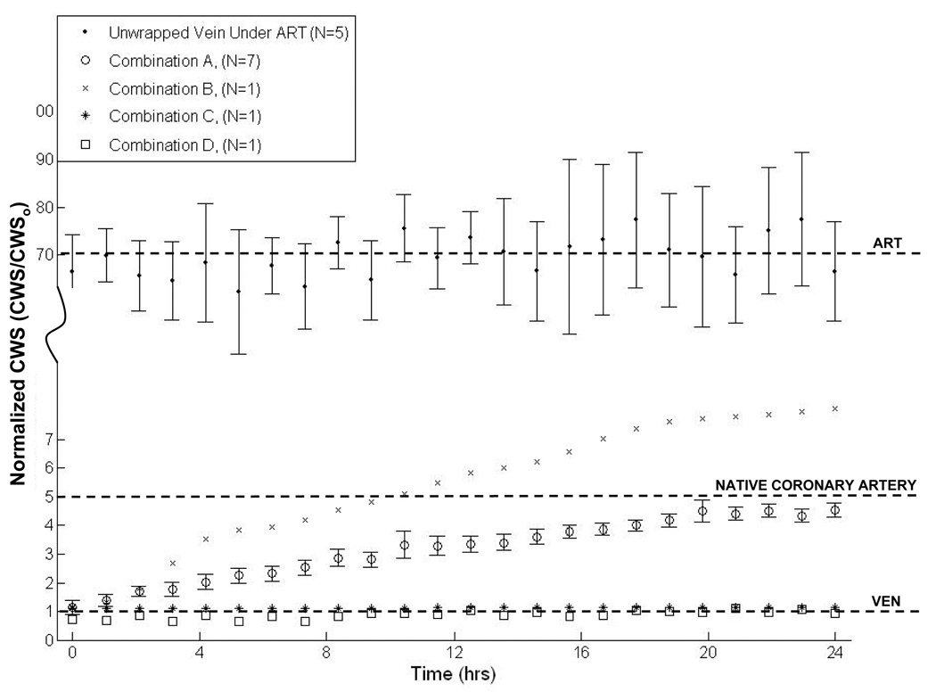
Arterial vein grafts (AVGs) often fail due to intimal hyperplasia, thrombosis, or accelerated atherosclerosis. Various approaches have been proposed to address AVG failure, including delivery of temporary mechanical support, many of which could be facilitated by perivascular placement of a biodegradable polymer wrap. The purpose of this work was to demonstrate that a polymer wrap can be applied to vein segments without compromising viability/function, and to demonstrate one potential application, i.e., gradually imposing the mid-wall circumferential wall stress (CWS) in wrapped veins exposed to arterial levels of pressure. Poly(ester urethane)urea, collagen, and elastin were combined in solution, and then electrospun onto freshly-excised porcine internal jugular vein segments. Tissue viability was assessed via Live/Dead staining for necrosis, and vasomotor challenge with epinephrine and sodium nitroprusside for functionality. Wrapped vein segments were also perfused for 24h within an ex vivo vascular perfusion system under arterial conditions (pressure = 120/80 mmHg; flow = 100 mL/min), and CWS was calculated every hour. Our results showed that the electrospinning process had no deleterious effects on tissue viability, and that the mid-wall CWS vs. time profile could be dictated through the composition and degradation of the electrospun wrap. This may have important clinical applications by enabling the engineering of an improved AVG.
Figures
Similar articles
-
Enhancement of tissue factor expression by vein segments exposed to coronary arterial hemodynamics.J Vasc Surg. 1998 Mar;27(3):521-7. doi: 10.1016/s0741-5214(98)70327-1. J Vasc Surg. 1998. PMID: 9546239
-
A biodegradable perivascular wrap for controlled, local and directed drug delivery.J Control Release. 2012 Jul 10;161(1):81-9. doi: 10.1016/j.jconrel.2012.04.029. Epub 2012 Apr 27. J Control Release. 2012. PMID: 22561340 Free PMC article.
-
Elasticity assessment of electrospun nanofibrous vascular grafts: a comparison with femoral ovine arteries.Mater Sci Eng C Mater Biol Appl. 2014 Dec;45:446-54. doi: 10.1016/j.msec.2014.09.016. Epub 2014 Sep 16. Mater Sci Eng C Mater Biol Appl. 2014. PMID: 25491850
-
Development of an in vitro model to study the response of saphenous vein endothelium to pulsatile arterial flow and circumferential deformation.Eur J Vasc Endovasc Surg. 1997 Jun;13(6):605-12. doi: 10.1016/s1078-5884(97)80071-8. Eur J Vasc Endovasc Surg. 1997. PMID: 9236715
-
Vein interposition cuffs decrease the intimal hyperplastic response of polytetrafluoroethylene bypass grafts.J Vasc Surg. 2000 Jan;31(1 Pt 1):69-83. doi: 10.1016/s0741-5214(00)70069-3. J Vasc Surg. 2000. PMID: 10642710
Cited by
-
Bioengineered human arterial equivalent and its applications from vascular graft to in vitro disease modeling.iScience. 2024 Oct 19;27(11):111215. doi: 10.1016/j.isci.2024.111215. eCollection 2024 Nov 15. iScience. 2024. PMID: 39555400 Free PMC article. Review.
-
The biomechanics and prevention of vein graft failure in coronary revascularization.Vessel Plus. 2023;7:31. doi: 10.20517/2574-1209.2023.97. Epub 2023 Dec 14. Vessel Plus. 2023. PMID: 39639997 Free PMC article.
-
Advances in preclinical surgical therapy of cardiovascular diseases.Int J Surg. 2024 Aug 1;110(8):4965-4975. doi: 10.1097/JS9.0000000000001534. Int J Surg. 2024. PMID: 38701509 Free PMC article. Review.
-
Correlation of tissue drug concentrations with in vivo magnetic resonance images of polymer drug depot around arteriovenous graft.J Control Release. 2010 Aug 17;146(1):23-30. doi: 10.1016/j.jconrel.2010.05.005. Epub 2010 May 8. J Control Release. 2010. PMID: 20457189 Free PMC article.
-
Biomimetic Approaches in Scaffold-Based Blood Vessel Tissue Engineering.Biomimetics (Basel). 2024 Jun 22;9(7):377. doi: 10.3390/biomimetics9070377. Biomimetics (Basel). 2024. PMID: 39056818 Free PMC article. Review.
References
-
- AHA. Heart disease and stroke statistics: 2006 update. Dallas, TX: American Heart Association; 2006.
-
- Feinglass J, Kaushik S, Handel D, Kosifas A, Martin G, Pearce WH. Peripheral bypass surgery and amputation. Arch. Surg. 2000;135:75. - PubMed
-
- Shears LL, Kibbe MR, Murdock AD, Billiar TR, Lizonova A, Kovesdi I, et al. Efficient inhibition of intimal hyperplasia by adenovirus-mediated inducible nitric oxide synthase gene transfer to rats and pigs in vivo. J Am Coll Surg. 1998;187(3):295–306. - PubMed
-
- Statistics, NCfH. Quarterly fact sheet: Monitoring health care in america. Hyattsville, MD: National Center for Health Statistics; 1996.
-
- Petrofski JA, Hata JA, Gehrig TR, Hanish SI, Williams ML, Thompson RB, et al. Gene delivery to aortocoronary saphenous vein grafts in a large animal model of intimal hyperplasia. J Thorac Cardiovasc Surg. 2004;127(1):27–33. - PubMed
Publication types
MeSH terms
Substances
Grants and funding
LinkOut - more resources
Full Text Sources
Other Literature Sources
